. 2018 Mar 28;13(3):e0194706.
doi: 10.1371/journal.pone.0194706. eCollection 2018.

Traction force microscopy of engineered cardiac tissues

Affiliations

Traction force microscopy of engineered cardiac tissues

Francesco Silvio Pasqualini et al. PLoS One. .

Abstract

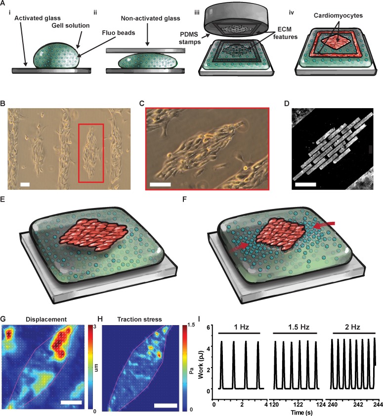
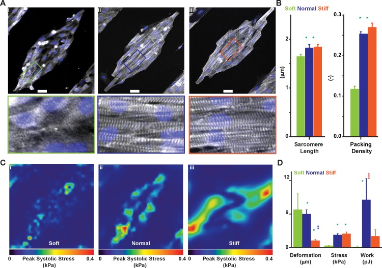
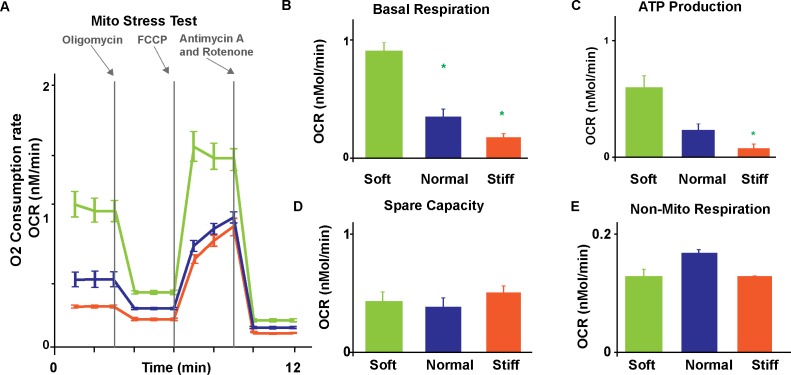
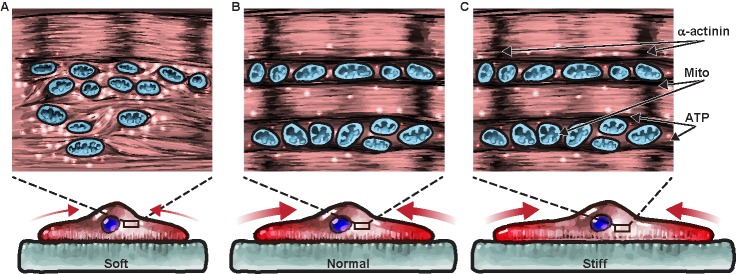
Cardiac tissue development and pathology have been shown to depend sensitively on microenvironmental mechanical factors, such as extracellular matrix stiffness, in both in vivo and in vitro systems. We present a novel quantitative approach to assess cardiac structure and function by extending the classical traction force microscopy technique to tissue-level preparations. Using this system, we investigated the relationship between contractile proficiency and metabolism in neonate rat ventricular myocytes (NRVM) cultured on gels with stiffness mimicking soft immature (1 kPa), normal healthy (13 kPa), and stiff diseased (90 kPa) cardiac microenvironments. We found that tissues engineered on the softest gels generated the least amount of stress and had the smallest work output. Conversely, cardiomyocytes in tissues engineered on healthy- and disease-mimicking gels generated significantly higher stresses, with the maximal contractile work measured in NRVM engineered on gels of normal stiffness. Interestingly, although tissues on soft gels exhibited poor stress generation and work production, their basal metabolic respiration rate was significantly more elevated than in other groups, suggesting a highly ineffective coupling between energy production and contractile work output. Our novel platform can thus be utilized to quantitatively assess the mechanotransduction pathways that initiate tissue-level structural and functional remodeling in response to substrate stiffness.


Conflict of interest statement

Competing Interests: The authors have declared that no competing interests exist.

Figures

Fig 1
Fig 1. Micropatterned cardiac tissues on soft gels for traction force microscopy.
(A) We cast PA gels (i) sandwiching the pre-polymer solution between activated and non-activated glass (ii) before stamping fibronectin (iii) to promote cell adhesion on the gel surface (iv). With this versatile method, we engineered neonate rat ventricular myocytes (NRVM) into diamond-shaped mini tissues (B) featuring cells aligned along the major axis of the diamond (C) thanks to a micro-contact printed brick-wall pattern of fibronectin (D). By tracking the displacement of fluorescent beads embedded in the in the soft gel during cell relaxation (E) and contraction (F), we used traction force microscopy to obtain displacement (G), and stress (H) maps at the tissue level. Importantly, since we could electrically stimulate the diamond-shaped tissues, we could measure displacement, stress, and contractile work as a function of beating frequency (I). Scale bars: 100 μm.
Fig 2
Fig 2. Contractile structure and function of cardiac tissues engineered on gels of varying stiffness.
Using α-actinin immunographs from NRVM engineered on soft (1 kPa, A-i), normal (13 kPa, A-ii), and stiff (90 kPa, A-iii) gels we assessed differences in sarcomere length (SL) and sarcomere packing density (SPD, B). Scale bar 50 μm. Further, we performed TFM on diamond-shaped NRVM tissues engineered on soft (C-i), normal (C-ii), and stiff (C-iii) gels (peak systolic stress shown) to calculate (D) maximum deformation, peak stress, and total contractile work (strain energy). Results are given as mean ± s.e.m. and N = 3, 4, and 7 for soft, normal, and stiff gels respectively. The symbol * implies significant differences (p < 0.05) compared with the group of the same color.
Fig 3
Fig 3. Metabolic differences in engineered cardiac tissues on gels of different stiffness.
(A) We measured oxygen consumption rates of NRVM cultured on soft (1 kPa), normal (13 kPa), and stiff (90 kPa) gels during a standard mitochondria stress test. We assessed basal respiration rate (B), ATP production (C), spare respiratory capacity (D), and nonmitochondrial respiration (E) for each substrate. Results are given as mean ± s.e.m. with sample size equal to 12, 15, and 22 for soft, normal, and stiff gels, respectively. The symbol * implies significant differences (p < 0.05) compared with the group of the same color.
Fig 4
Fig 4. Cardiomyocytes operate optimally on substrates of physiological stiffness possibly due to a link between metabolism and contractile structure and function.
Based on our data, we speculated that substrate stiffness might regulate the balance of energy production and utilization in cardiac tissues as follows. Cardiomyocytes on soft gels (A) need abundant ATP (white spheres) derived by mitochondria (orange organelles) to promote the contraction of sarcomeres (Z-disks in black) via cross-bridge cycling (brown lines). This might lead to very inefficient mechanical work as only a small stress (red arrows) is required to deform the soft gel substantially. Conversely, NRVM cultured on normal (B) and stiff (C) substrates have well organized contractile cytoskeletons and need a limited amount of ATP to fuel sarcomere contraction. Since the same amount of contractile force causes a smaller displacement of stiffer gels, the product between force and displacement (work) is maximum on gels of physiological stiffness.

References

    1. Chabiniok R, Wang VY, Hadjicharalambous M, Asner L, Lee J, Sermesant M, et al. Multiphysics and multiscale modelling, data-model fusion and integration of organ physiology in the clinic: ventricular cardiac mechanics. Interface focus. 2016;6(2):20150083 doi: 10.1098/rsfs.2015.0083 - DOI - PMC - PubMed
    1. Parker KK, Ingber DE. Extracellular matrix, mechanotransduction and structural hierarchies in heart tissue engineering. Philosophical transactions of the Royal Society of London Series B, Biological sciences. 2007;362(1484):1267–79. doi: 10.1098/rstb.2007.2114 - DOI - PMC - PubMed
    1. Sheehy SP, Grosberg A, Qin P, Behm DJ, Ferrier JP, Eagleson MA, et al. Toward improved myocardial maturity in an organ-on-chip platform with immature cardiac myocytes. Experimental biology and medicine (Maywood, NJ). 2017:1535370217701006. - PMC - PubMed
    1. Sheehy SP, Pasqualini F, Grosberg A, Park SJ, Aratyn-Schaus Y, Parker KK. Quality metrics for stem cell-derived cardiac myocytes. Stem cell reports. 2014;2(3):282–94. doi: 10.1016/j.stemcr.2014.01.015 - DOI - PMC - PubMed
    1. Aratyn-Schaus Y, Pasqualini FS, Yuan H, McCain ML, Ye GJ, Sheehy SP, et al. Coupling primary and stem cell-derived cardiomyocytes in an in vitro model of cardiac cell therapy. The Journal of cell biology. 2016;212(4):389–97. doi: 10.1083/jcb.201508026 - DOI - PMC - PubMed

Publication types

LinkOut - more resources