. 2018 Mar 28;13(3):e0195108.
doi: 10.1371/journal.pone.0195108. eCollection 2018.

The neurochemistry and morphology of functionally identified corneal polymodal nociceptors and cold thermoreceptors

Affiliations

The neurochemistry and morphology of functionally identified corneal polymodal nociceptors and cold thermoreceptors

Abdulhakeem S Alamri et al. PLoS One. .

Abstract

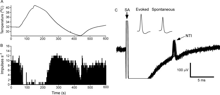
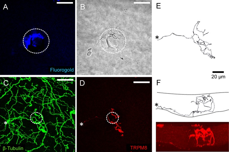
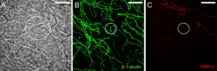
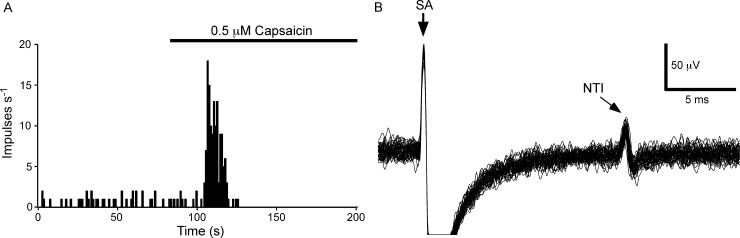
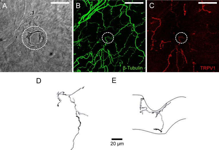
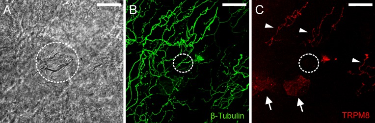
It is generally believed that the unencapsulated sensory nerve terminals of modality specific C- and Aδ-neurons lack structural specialization. Here we determined the morphology of functionally defined polymodal receptors and cold thermoreceptors in the guinea pig corneal epithelium. Polymodal receptors and cold thermoreceptors were identified by extracellular recording at the surface of the corneal epithelium. After marking the recording sites, corneas were processed to reveal immunoreactivity for the transient receptor potential channels TRPV1 (transient receptor potential cation channel, subfamily V, member 1) or TPRM8 (transient receptor potential cation channel subfamily M member 8). Polymodal receptor nerve terminals (n = 6) were TRPV1-immunoreactive and derived from an axon that ascended from the sub-basal plexus to the squamous cell layer where it branched into fibers that ran parallel to the corneal surface and terminated with small bulbar endings (ramifying endings). Cold thermoreceptor nerve terminals were TRPM8-immunoreactive (n = 6) and originated from an axon that branched as it ascended through the wing cell and squamous cell layers and terminated with large bulbar endings (complex endings). These findings indicate that modality specific corneal sensory neurons with unencapsulated nerve endings have distinct nerve terminal morphologies that are likely to relate to their function.


Conflict of interest statement

Competing Interests: The authors have declared that no competing interests exist.

Figures

Fig 1
Fig 1. The electrical activity recorded from a cold thermoreceptor nerve terminal at the corneal surface.
A and B, show the temperature of the solution superfusing the cornea (A) and the frequency of nerve terminal impulse (NTI) discharge (B). During heating and cooling the frequency of NTIs was decreased and increased, respectively. C, overlaid traces recorded during stimulation of the ciliary nerves with a train of 25 pulses at 1 Hz. At this recording site electrical stimulation evoked a single stimulus locked NTI. The insets in C show averages of the electrically evoked and spontaneous NTIs. The smaller amplitude of the spontaneous NTIs indicates they are likely to be initiated very close to site of recording (see [25]). In C, the stimulus artifact (SA) is indicated and during the flat line immediately following the SA the signal was out of the analogue-to-digital recording range.
Fig 2
Fig 2. Immuno-labeling for TRPM8 at the cold thermoreceptor recording site where the electrical activity shown in Fig 1 was recorded.
A, fluoro-gold fluorescence within the white dotted circle identifies the approximate location of the recording site. B, bright-field image showing within the white dotted circle the mark left on the corneal surface by the rim of the recording electrode. C, all axons in the field of view were revealed by β-tubulin III-IR (green). D, a TRPM8-IR (red) nerve terminal was located at the recording site. In C and D, the white dotted circle approximates dimensions of the electrode tip at the recording site revealed in B. The scale bar in A–D = 50 μm. E and F, show a 3-dimensional reconstruction of the TRPM8-IR nerve terminal viewed from the front (E) and side (F). Panel F shows an orthogonal projection of the image shown in D. In C–F, the asterisk marks the site where the parent axon of the TRPM8-IR nerve terminal enters into the epithelium through Bowman’s membrane. The dashed lines in F approximate the area occupied by the corneal epithelium.
Fig 3
Fig 3. Immuno-labeling for TRPV1 at a cold thermoreceptor recording site.
A, bright-field image showing within the white dotted circle the mark left on the corneal surface by the rim of the recording electrode. B, all axons in the field of view were revealed by β-tubulin III-IR (green). C, while TRPV1-IR axons (red) were observed within the field of view, those enclosed by the recording electrode were not immunoreactive for this protein. In B and C, the dotted circle approximates the dimensions of the electrode tip at the recording site revealed in A. The scale bar in A–C = 50 μm.
Fig 4
Fig 4. The electrical activity recorded from a capsaicin-sensitive nerve terminal at the corneal surface.
A, the frequency of NTI discharge before and during application of capsaicin (0.5 μM). In this receptor, there was a low level of ongoing NTI activity that was markedly increased by capsaicin. B, overlaid traces recorded during stimulation of the ciliary nerves with a train of 25 pulses at 1 Hz. At this recording site electrical stimulation evoked a single stimulus locked NTI. In B, the stimulus artifact (SA) is indicated and during the flat line immediately following the SA the signal was out of the analogue-to-digital recording range.
Fig 5
Fig 5. Immuno-labeling for TRPV1 at the polymodal receptor recording site where the electrical activity shown in Fig 4 was recorded.
A, bright-field image showing within the white dotted circle the mark left on the corneal surface by the rim of the recording electrode. B, all axons in the field of view were revealed by β-tubulin III-IR (green). C, a TRPV1-IR (red) nerve terminal was located at the recording site. In B and C, the dotted circle approximates the dimensions of the electrode tip at the recording site revealed in A. The scale bar in A–C = 50 μm. D and E, show a 3-dimensional reconstruction of the TRPV1-IR nerve terminal from front (D) and side (E) views. The dashed lines in E approximate the boundaries of the corneal epithelium. The indentation of the epithelium was produced by the recording electrode.
Fig 6
Fig 6. Immuno-labeling for TRPM8 at a polymodal receptor recording site.
A, bright-field image showing within the white dotted circle the mark left on the corneal surface by the rim of the recording electrode. B, all axons in the field of view were revealed by β-tubulin III-IR (green). C, while TRPM8-IR axons (red) were located above and to the right of the recording site (indicated by arrowheads), those enclosed by the recording electrode were not immunoreactive for this protein. In C, there are small patches of fluorescence located on the surface of the cornea epithelium (arrows). These are an artifact produced by light reflecting off the surface of the epithelium. In B and C, the dotted circle approximates the dimensions of the electrode tip at the recording site revealed in A. The scale bar in A–C = 50 μm.

References

    1. Owens DM, Lumpkin EA. Diversification and specialization of touch receptors in skin. Cold Spring Harb Perspect Med. 2014; 4(6). doi: 10.1101/cshperspect.a013656 - DOI - PMC - PubMed
    1. Zimmerman A, Bai L, Ginty DD. The gentle touch receptors of mammalian skin. Science. 2014;346(6212):950–954. doi: 10.1126/science.1254229 - DOI - PMC - PubMed
    1. Hensel H, Andres KH, von During M. Structure and function of cold receptors. Pflugers Archiv: Eur J Physiol. 1974; 352: 1–10. - PubMed
    1. Heppelmann B, Gallar J, Trost B, Schmidt RF, Belmonte C. Three-dimensional reconstruction of scleral cold thermoreceptors of the cat eye. J Comp Neurol. 2001; 44: 148–154. - PubMed
    1. Li L, Ginty DD. The structure and organization of lanceolate mechanosensory complexes at mouse hair follicles. Elife. 2014; 3: e01901 doi: 10.7554/eLife.01901 - DOI - PMC - PubMed

Publication types

Substances

LinkOut - more resources