. 2018 Mar 28;14(3):e1006964.
doi: 10.1371/journal.ppat.1006964. eCollection 2018 Mar.

Molecularly barcoded Zika virus libraries to probe in vivo evolutionary dynamics

Affiliations

Molecularly barcoded Zika virus libraries to probe in vivo evolutionary dynamics

Matthew T Aliota et al. PLoS Pathog. .

Abstract

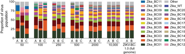
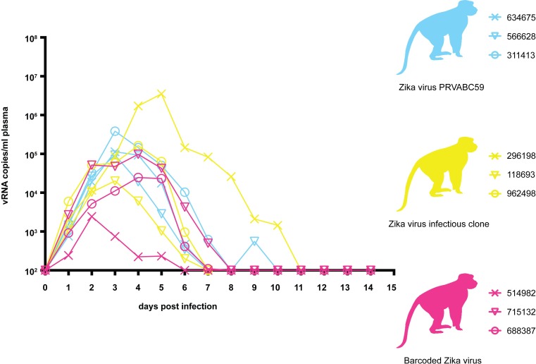
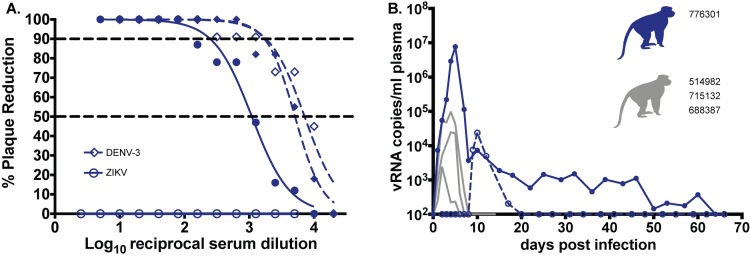
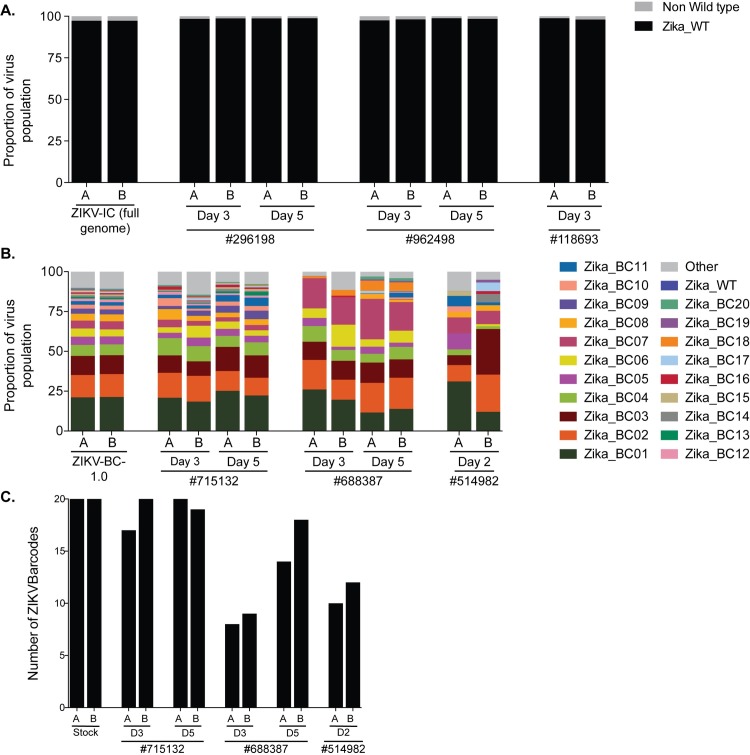
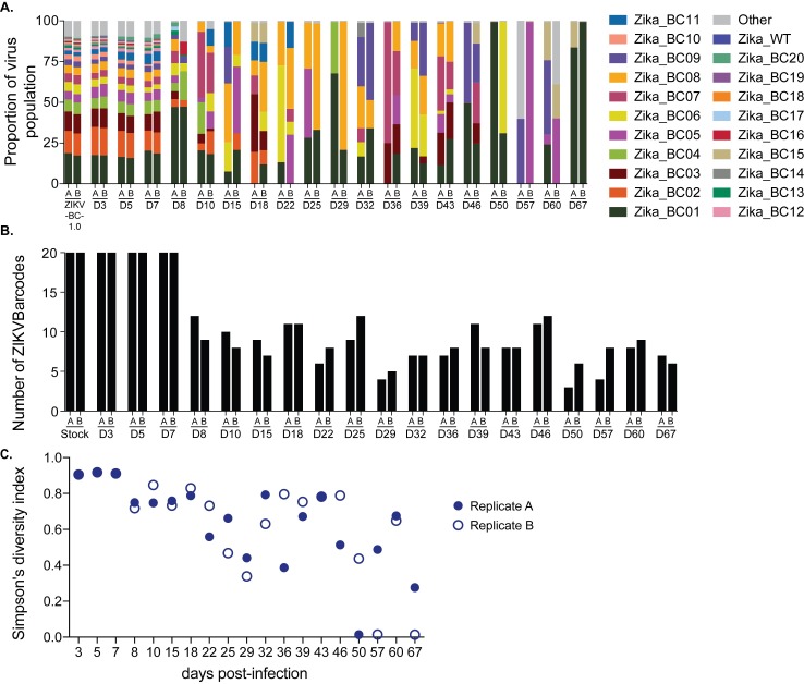
Defining the complex dynamics of Zika virus (ZIKV) infection in pregnancy and during transmission between vertebrate hosts and mosquito vectors is critical for a thorough understanding of viral transmission, pathogenesis, immune evasion, and potential reservoir establishment. Within-host viral diversity in ZIKV infection is low, which makes it difficult to evaluate infection dynamics. To overcome this biological hurdle, we constructed a molecularly barcoded ZIKV. This virus stock consists of a "synthetic swarm" whose members are genetically identical except for a run of eight consecutive degenerate codons, which creates approximately 64,000 theoretical nucleotide combinations that all encode the same amino acids. Deep sequencing this region of the ZIKV genome enables counting of individual barcodes to quantify the number and relative proportions of viral lineages present within a host. Here we used these molecularly barcoded ZIKV variants to study the dynamics of ZIKV infection in pregnant and non-pregnant macaques as well as during mosquito infection/transmission. The barcoded virus had no discernible fitness defects in vivo, and the proportions of individual barcoded virus templates remained stable throughout the duration of acute plasma viremia. ZIKV RNA also was detected in maternal plasma from a pregnant animal infected with barcoded virus for 67 days. The complexity of the virus population declined precipitously 8 days following infection of the dam, consistent with the timing of typical resolution of ZIKV in non-pregnant macaques and remained low for the subsequent duration of viremia. Our approach showed that synthetic swarm viruses can be used to probe the composition of ZIKV populations over time in vivo to understand vertical transmission, persistent reservoirs, bottlenecks, and evolutionary dynamics.


Conflict of interest statement

The authors have declared that no competing interests exist.

Figures

Fig 1
Fig 1. Sequencing of the titrated ZIKV-BC-1.0 stock.
Dilutions of ZIKV-BC-1.0 were reverse-transcribed, PCR-amplified with a single primer pair, and sequenced, as described in the materials and methods. The number of vRNA templates that were used for each cDNA synthesis reaction is shown below the graph. The theoretical number of cDNA molecules used in each PCR reaction is shown in S5 Table. Each dilution was sequenced in triplicate, with individual replicates labeled A, B, and C. As a comparison, data for ZIKV-BC-1.0 that was collected by the multiplex PCR approach (Table 1) are shown on the far right. The frequency of each authentic barcode was enumerated and are shown. The frequency of the wild type sequence in the region of the barcode is shown as Zika_WT. The frequency of any sequence in the region of the barcode that was not considered authentic is listed as ‘Other.’
Fig 2
Fig 2. Longitudinal detection of Zika vRNA in plasma from animals inoculated with ZIKV-PR (blue), ZIKV-IC (yellow), or ZIKV-BC-1.0 (magenta).
Zika vRNA copies per ml blood plasma. The y-axis crosses the x-axis at the limit of quantification of the qRT-PCR assay (100 vRNA copies/ml).
Fig 3
Fig 3. Maternal Zika vRNA loads and neutralization curves.
A.) Neutralization by ZIKV and DENV-3 immune sera from a pregnant ZIKV-BC-1.0-infected macaque. Immune sera from a macaque infected with ZIKV-BC-1.0 during the first trimester of pregnancy was tested for its capacity to neutralize DENV-3 (blue dashes) and ZIKV-PR (blue). Infection was measured by plaque reduction neutralization test (PRNT) and is expressed relative to the infectivity of ZIKV-PR in the absence of serum. The concentration of sera indicated on the x-axis is expressed as log10 (dilution factor of serum). The EC90 and EC50, estimated by non-linear regression analysis, are also indicated by a dashed line. Neutralization curves for each virus (ZIKV, solid blue; DENV-3, dashed blue) at 0 (open symbols) and 28 (closed symbols) dpi are shown. B.) Zika vRNA copies per ml blood plasma (solid lines) or urine (dashed line). Blue tracings represent the animal infected with ZIKV-BC-1.0 at 35 days gestation. The day of gestation is estimated +/- 2 days. Grey tracings represent viremia in nonpregnant/male rhesus monkeys infected with the identical dose of ZIKV-BC-1.0 (Fig 2). The y-axis crosses the x-axis at the limit of quantification of the qRT-PCR assay (100 vRNA copies/ml).
Fig 4
Fig 4. Maternal and fetal histopathology analysis: Hematoxylin and eosin (H&E) staining of selected tissues.
A.) Fetal spleen: increased neutrophils (arrows) throughout splenic sinusoids of the red pulp. B.) Fetal inguinal lymph node: increased neutrophils (arrows) throughout central sinusoids. C.) Placental artery, maternal side: numerous neutrophils (arrow) within the vascular lumen, with neutrophilic inflammation extending into the vascular wall (W). D.) Maternal decidua: normal for gestational age remodeled artery (N) and a persistently muscularized artery (M). Numerous plasma cells (P) within the decidual stroma. Scale bar = 100 µm.
Fig 5
Fig 5. Sequencing of ZIKV-BC-1.0 and ZIKV-IC isolated from nonpregnant rhesus macaques.
ZIKV RNA was isolated from plasma at the indicated time points from each of the animals infected with A.) ZIKV-IC or B.) ZIKV-BC-1.0. Viral RNA was reverse transcribed and then multiplex PCR was performed as described in materials and methods. PCR products were tagged and sequenced. A.) The sequence mapping to the region containing the molecular barcode was interrogated for ZIKV-IC, and the frequency of wild type and non-wild type ZIKV sequences are shown. The theoretical number of cDNA molecules used in each PCR reaction is shown in Table 2. Each sample was sequenced in duplicate, as labeled by A and B. B.) The frequency of each barcode in the population is shown for ZIKV-BC-1.0 and the nonpregnant animals infected with ZIKV-BC-1.0. The frequency of the wild type sequence in the region of the barcode is shown as Zika_WT. The frequency of any sequence in the region of the barcode that was not considered authentic is listed as ‘Other.’ C.) The number of authentic barcodes detected in the three nonpregnant animals and the stock were counted. The data for each individual replicate are shown.
Fig 6
Fig 6. Sequencing of the molecular barcode isolated from pregnant animal 776301.
Viral RNA was isolated from animal 776301 at the indicated time points. The theoretical number of cDNA molecules used in each PCR reaction is shown in Table 3. For each sample, a single preparation of cDNA was made and then split into two separate PCR reactions with primer set A (178 bp) and B (131bp). PCR products were tagged and sequenced. A.) The frequency of each barcode in the population is shown for ZIKV-BC-1.0 inoculum and the animal 776301. The frequency of the wild type sequence in the region of the barcode is shown as Zika_WT. The frequency of any sequence in the region of the barcode that was not considered authentic is listed as ‘Other.’ B.) The number of authentic barcodes detected in animal 776301 were counted for each sample and the data for each individual replicate are shown. C.) Average genetic complexity at the barcode positions measured by Simpson’s diversity index. Closed symbols represent Simpson’s diversity index in replicate A samples and open symbols represent Simpson’s diversity index in replicate B.
Fig 7
Fig 7. Sequencing of the molecular barcode isolated from mosquito #27 that fed on 776301.
Viral RNA that was isolated from the saliva, body, and legs of mosquito #27 at day 25 post feeding was converted into cDNA and then split into two separate PCR reactions with primer set A (178 bp) and B (131 bp). The theoretical number of cDNA molecules used in each PCR reaction is shown in Table 5. PCR products were tagged and sequenced. The frequency of each barcode detected was quantified. Mosquito #27 fed on 776301 on day 4, so the frequencies of barcodes detected in the plasma of 776301 from Fig 6 are shown. The frequency of any sequence in the region of the barcode that was not considered authentic is listed as ‘Other.’

References

    1. Reynolds MR, Jones AM, Petersen EE, Lee EH, Rice ME, Bingham A et al. (2017) Vital Signs: Update on Zika Virus-Associated Birth Defects and Evaluation of All U.S. Infants with Congenital Zika Virus Exposure—U.S. Zika Pregnancy Registry, 2016. MMWR Morb Mortal Wkly Rep 66: 366–373. doi: 10.15585/mmwr.mm6613e1 - DOI - PMC - PubMed
    1. Nguyen SM, Antony KM, Dudley DM, Kohn S, Simmons HA, Wolfe B et al. (2017) Highly efficient maternal-fetal Zika virus transmission in pregnant rhesus macaques. PLoS Pathog 13: e1006378 doi: 10.1371/journal.ppat.1006378 - DOI - PMC - PubMed
    1. Rosenberg K (2017) Zika Virus can Persist in Body Fluids for Prolonged Periods. Am J Nurs 117: 71. - PubMed
    1. Musso D, Roche C, Robin E, Nhan T, Teissier A, Cao-Lormeau VM (2015) Potential sexual transmission of Zika virus. Emerg Infect Dis 21: 359–361. doi: 10.3201/eid2102.141363 - DOI - PMC - PubMed
    1. Aid M, Abbink P, Larocca RA, Boyd M, Nityanandam R, Nanayakkara O et al. (2017) Zika Virus Persistence in the Central Nervous System and Lymph Nodes of Rhesus Monkeys. Cell 169: 610–620.e14. doi: 10.1016/j.cell.2017.04.008 - DOI - PMC - PubMed

Publication types