. 2018 Mar 27;22(13):3587-3597.
doi: 10.1016/j.celrep.2018.02.102.

Leucine Zipper-Bearing Kinase Is a Critical Regulator of Astrocyte Reactivity in the Adult Mammalian CNS

Affiliations

Leucine Zipper-Bearing Kinase Is a Critical Regulator of Astrocyte Reactivity in the Adult Mammalian CNS

Meifan Chen et al. Cell Rep. .

Abstract

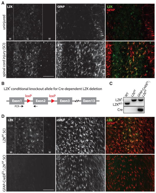
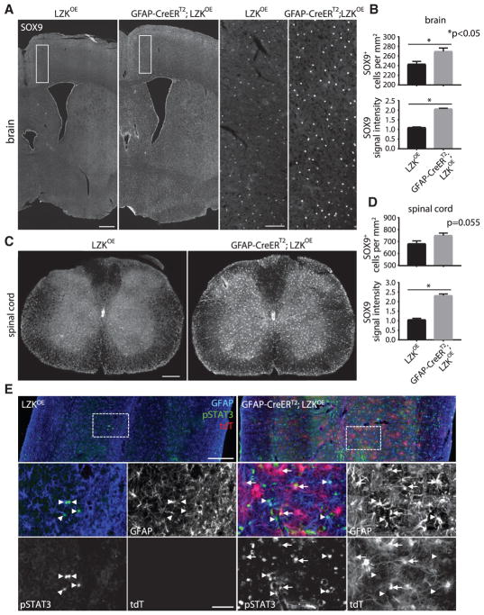
Reactive astrocytes influence post-injury recovery, repair, and pathogenesis of the mammalian CNS. Much of the regulation of astrocyte reactivity, however, remains to be understood. Using genetic loss and gain-of-function analyses in vivo, we show that the conserved MAP3K13 (also known as leucine zipper-bearing kinase [LZK]) promotes astrocyte reactivity and glial scar formation after CNS injury. Inducible LZK gene deletion in astrocytes of adult mice reduced astrogliosis and impaired glial scar formation, resulting in increased lesion size after spinal cord injury. Conversely, LZK overexpression in astrocytes enhanced astrogliosis and reduced lesion size. Remarkably, in the absence of injury, LZK overexpression alone induced widespread astrogliosis in the CNS and upregulated astrogliosis activators pSTAT3 and SOX9. The identification of LZK as a critical cell-intrinsic regulator of astrocyte reactivity expands our understanding of the multicellular response to CNS injury and disease, with broad translational implications for neural repair.

Keywords: CNS injury; LZK; MAP3K; SOX9; STAT3; astroglial reactivity; astrogliosis; glial scar; reactive astrocytes; spinal cord injury.


Conflict of interest statement

DECLARATION OF INTERESTS

The authors declare no competing interests.

Figures

Figure 1
Figure 1. Injury-Induced Leucine Zipper-Bearing Kinase Expression in Astrocytes and Conditional Gene Deletion
(A) Representative images of endogenous LZK and glial fibrillary astrocyte protein (GFAP) immunostaining in the spinal cords of uninjured or injured wild-type (WT) mice (14 days after spinal cord injury [SCI]), taken 0.5–1 mm from the injury site on horizontal sections. (B) Diagram of the LZK conditional knockout allele (LZKf). Cre-mediated excision of exon 2 would result in a frameshift and thus a null allele. Black arrows mark the positions of PCR primers for genotyping. (C) Genomic PCR genotyping of WT, LZKf/f, and GFAP-CreERT2;LZKf/f mice. (D) Immunostaining of endogenous LZK and GFAP in the spinal cords of tamoxifen-pretreated LZKf/f control and GFAP-CreERT2;LZKf/f mice 14 days after spinal cord injury (SCI), with images taken 0.5–1 mm from the injury site. Dotted lines demarcate white (W) matter and gray (G) matter. Scale bars represent 100 μm. See also Figure S1.
Figure 2
Figure 2. LZK Deletion in Adult Astrocytes Impaired Astrogliosis 14 Days after Spinal Cord Injury
(A) Representative images of GFAP and DAPI nuclear staining centered at the spinal cord injury site from tamoxifen-pretreated LZKf/f control and GFAP-CreERT2;LZKf/f mice. Note that the GFAP lesion core is enveloped by GFAP+ astrocytes and their processes. Areas within the white boxes are shown in high magnification (rightmost panels) to illustrate astrocytic processes parallel (arrows) to the lesion border in control mice but perpendicular (arrowheads) in astrocytic LZK knockout mice. Figures are composites of smaller microscopy images. Scale bar represents 200 μm (low magnification) and 100 μm (high magnification). (B) Quantification of lesion size in LZKf/f control versus GFAP-CreERT2;LZKf/f mice. n = 8 mice per genotype, *p < 0.05 by unpaired parametric t test. (C) GFAP immunofluorescence intensity in the injured spinal cords of LZKf/f control versus GFAP-CreERT2;LZKf/f mice in 9 zones (each the width of the spinal cord and length of 250 μm). Zone 1 starts at the lesion border, followed by the other zones placed sequentially away from the injury site and immediately adjacent to one another, as adapted from Wanner et al., (2013). n = 8 mice per genotype, *p < 0.05 by two-way ANOVA followed by post hoc multiple t test between groups for each zone. Error bar represents SEM.
Figure 3
Figure 3. LZK Deletion in Adult Astrocytes Reduced Astrogliosis in the Injured Spinal Cord as Assessed by Vimentin Expression and Astrocyte Proliferation
(A) Representative images of vimentin and DAPI staining at the spinal cord injury site of tamoxifen-treated LZKf/f control and GFAP-CreERT2;LZKf/f mice 14 days post-injury (dpi). Astrocytes strongly expressing vimentin enclose the lesion core. Areas within white boxes are shown in high magnification (rightmost panels) to illustrate astrocytic processes parallel (arrows) and perpendicular (arrowheads) to the lesion border in control mice and mice lacking astrocytic LZK, respectively. Figures are composites of smaller microscopy images. (B) Representative images of DAPI, SOX9, and Ki67 co-immunofluorescence staining within 250 μm of the spinal cord injury site of tamoxifen-treated LZKf/f control and GFAP-CreERT2;LZKf/f mice at 7 dpi. (C) Quantification of vimentin immunofluorescence intensity at and within 250 μm of the lesion border in control versus astrocytic LZK knockout mice 14 dpi. n = 4 per genotype, *p < 0.05 by two-tailed unpaired parametric t test. (D) Quantification of the numbers of proliferating astrocytes by Ki67+SOX9+ co-labeling within 250 μm of spinal cord injury site in control versus astrocytic LZK knockout mice 7 dpi. n = 3 per genotype, *p < 0.05 by two-tailed unpaired parametric t test. Scale bars, 200 μm (A, low magnification), 100 μm (A, high magnification, rightmost panels), and 50 μm (B). See also Figure S2.
Figure 4
Figure 4. LZK Overexpression in Adult Astrocytes Enhanced Astrogliosis and Reduced Lesion Size 14 dpi
(A) Diagram of the LZK conditional overexpression transgene (LZKOE). LZKOE has two loxP sites flanking a STOP cassette upstream of an LZK-T2A-tdTomato (tdT) fusion gene. Cre-mediated excision of STOP would activate LZK-T2A-tdT, leading to overexpression of LZK and the associated fluorescent reporter tdT. Black arrows mark the positions of PCR primers for genotyping. (B) Genomic PCR genotyping of WT, LZKOE, and GFAP-CreERT2;LZKOE mice. (C) Representative images taken from perilesional area (0.5–1 mm from the injury site), showing that tdT activation was associated with LZK overexpression, GFAP upregulation, and astrocyte hypertrophy in tamoxifen-pretreated GFAP-CreERT2;LZKOE mice as compared with LZKOE control mice. Note LZK and GFAP co-localization in the merged panels. (D) Representative images of GFAP immunostaining, tdT direct fluorescence, and DAPI nuclear staining centered at the injury site in the spinal cords of tamoxifen-pretreated LZKOE control mice and GFAP-CreERT2;LZKOE mice. Areas within the white boxes are shown in high magnification (rightmost panels) to illustrate the lesion borders lined by GFAP+ astrocytes. Note the presence of tdT+ astrocytes and the more compact lesion in the GFAP-CreERT2;LZKOE mouse. Figures are composites of smaller microscopy images. Scale bar represents 250 μm (low magnification), 100 μm (high magnification). (E) Quantification of lesion size in LZKOE control versus GFAP-CreERT2;LZKOE mice. n = 5 for LZKOE mice; n = 3 for GFAP-CreERT2;LZKOE mice; p = 0.08 by unpaired parametric t test. See Figure S3 for an independent replicate experiment by a second surgeon. (F) GFAP immunofluorescence intensity in the injured spinal cords of LZKf/f control versus GFAP-CreERT2;LZKf/f mice in 9 zones defined similarly in Figure 2C. Same numbers of mice as in (E); *p < 0.05 by two-way ANOVA followed by post hoc multiple t test between groups for each zone. Error bar represents SEM. See also Figure S3.
Figure 5
Figure 5. LZK Overexpression in Adult Astrocytes Induced Widespread Astrogliosis in the Absense of Injury as Assessed by GFAP Immunoreactivity
(A and B) Representative images of GFAP immunostaining and tdT direct fluorescence on horizontal (A) and transverse (B) spinal cord sections from tamoxifen-treated, uninjured LZKOE control and GFAP-CreERT2;LZKOE mice. (C) Quantification of GFAP immunofluorescence intensity on horizontal spinal cord sections from LZKOE control versus GFAP-CreERT2;LZKOE mice after tamoxifen treatment. n = 3 mice per genotype, *p < 0.05 by unpaired parametric t test. (D) Higher magnification images of LZK, tdT, and GFAP signals on horizontal spinal cord sections from tamoxifen-treated, uninjured mice, showing co-localization of LZK/GFAP and noting similar patterns of LZK with tdT direct fluorescence. Dotted lines demarcate white (W) matter and gray (G) matter. (E) LZK, GFAP immunostaining, and tdT direct fluorescence in the brains of tamoxifen-treated LZKOE control and GFAP-CreERT2;LZKOE mice. (F) Quantification of GFAP immunofluorescence intensity in the cerebral cortices of tamoxifen-treated LZKOE versus GFAP-CreERT2;LZKOE mice. n = 3 mice per genotype, *p < 0.05 by unpaired parametric t test. Error bar represents SEM. Scale bars, 250 μm (A and B), 100 μm (D), and 1 mm (E). See also Figures S4 and S5.
Figure 6
Figure 6. LZK Overexpression in Adult Astrocytes Induced Widespread Astrogliosis in the Absence of Injury as Assessed by Vimentin Immunoreactivity and Astrocyte Proliferation
(A and B) Vimentin, GFAP immunostaining, and tdT direct fluorescence on coronal sections from the hippocampal region of the brains (A) and horizontal spinal cord sections (B) of LZKOE control and GFAP-CreERT2;LZKOE mice after tamoxifen treatment. Note the similarly upregulated vimentin and GFAP immunoreactivity in GFAP-CreERT2;LZKOE mice. (C) Ki67, GFAP immunostaining, and tdT direct fluorescence in the cerebral cortices (brain) and spinal cord gray matter from LZKOE control and GFAP-CreERT2;LZKOE mice. (D) Quantification of Ki67+GFAP+ cell numbers in the cerebral cortices (brain) and spinal cord gray matter of LZKOE control and GFAP-CreERT2;LZKOE mice. n = 3 per group, p value determined by unpaired parametric t test. Error bar represents SEM. Scale bars 250 μm (A and B) and 50 μm (C). (A) and (B) are composites of smaller microscopy images.
Figure 7
Figure 7. LZK Positively Regulates SOX9 and pSTAT3 in Adult Astrocytes In Vivo
(A) Immunofluorescence staining of SOX9 on coronal brain sections from LZKOE control and GFAP-CreERT2;LZKOE mice. Lower-magnification images are shown in the left panels, with cerebral cortical areas within the white boxes shown in high magnification in the right panels. Note the higher levels of SOX9 immunoreactivity in GFAP-CreERT2;LZKOE mice. (B) Quantification of SOX9+ cell number and SOX9 immunofluorescence intensity in the cerebral cortices of LZKOE control and GFAP-CreERT2;LZKOE mice. n = 3 per group, p value determined by unpaired parametric t test. (C) Immunofluorescence staining of SOX9 on transverse spinal cord sections. Note the higher levels of SOX9 immunoreactivity in GFAP-CreERT2;LZKOE mice. (D) Quantification of SOX9+ cell number and SOX9 immunofluorescence intensity in the spinal cord gray matter of LZKOE control and GFAP-CreERT2;LZKOE mice. n = 3 per group, p value determined by unpaired parametric t test. (E) Immunofluorescence detection of GFAP, pSTAT3, and tdT on horizontal spinal cord sections of LZKOE control (left panels) and GFAP-CreERT2;LZKOE mice (right panels). Arrows represent tdT/pSTAT3 co-labeled cells; arrowheads represent GFAP/pSTAT3 co-labeled cells but without tdT co-labeling. Quantification of these data (shown in Figures S3D and S3E) indicates that LZK overexpression in adult astrocytes increased pSTAT3+ cells/astrocytes in the uninjured spinal cord. Scale bars, 500 μm (A, low magnification), 100 μm (A, high magnification), 250 μm (C), 200 μm (E, low magnification), 50 μm (E, high magnification). (A), (C), and (E) are composites of smaller microscopy images.

References

    1. Anderson MA, Burda JE, Ren Y, Ao Y, O’Shea TM, Kawaguchi R, Coppola G, Khakh BS, Deming TJ, Sofroniew MV. Astrocyte scar formation aids central nervous system axon regeneration. Nature. 2016;532:195–200. - PMC - PubMed
    1. Ben Haim L, Rowitch DH. Functional diversity of astrocytes in neural circuit regulation. Nat Rev Neurosci. 2017;18:31–41. - PubMed
    1. Bradbury EJ, Moon LD, Popat RJ, King VR, Bennett GS, Patel PN, Fawcett JW, McMahon SB. Chondroitinase ABC promotes functional recovery after spinal cord injury. Nature. 2002;416:636–640. - PubMed
    1. Brambilla R, Bracchi-Ricard V, Hu WH, Frydel B, Bramwell A, Karmally S, Green EJ, Bethea JR. Inhibition of astroglial nuclear factor kappaB reduces inflammation and improves functional recovery after spinal cord injury. J Exp Med. 2005;202:145–156. - PMC - PubMed
    1. Burda JE, Sofroniew MV. Reactive gliosis and the multicellular response to CNS damage and disease. Neuron. 2014;81:229–248. - PMC - PubMed

Publication types