. 2018 Mar 28;25(1):28.
doi: 10.1186/s12929-018-0432-6.

Novel microtubule inhibitor MPT0B098 inhibits hypoxia-induced epithelial-to-mesenchymal transition in head and neck squamous cell carcinoma

Affiliations

Novel microtubule inhibitor MPT0B098 inhibits hypoxia-induced epithelial-to-mesenchymal transition in head and neck squamous cell carcinoma

I-Ting Tsai et al. J Biomed Sci. .

Abstract

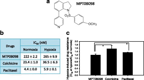
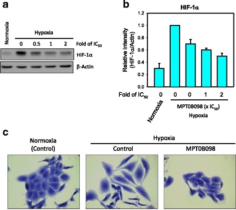
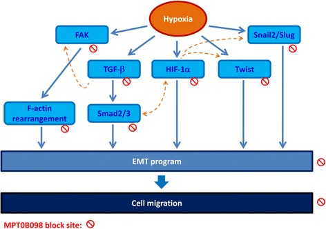
Background: Tumor hypoxia-induced epithelial-mesenchymal transition (EMT) is critical in promoting cancer metastasis. We recently discovered a novel microtubule inhibitor, MPT0B098, that employs a novel antitumor mechanism. It destabilizes hypoxia-inducible factor (HIF)-1α mRNA by blocking the function of human antigen R. Thus, we proposed that MPT0B098 modulates hypoxia-induced EMT.

Methods: In vitro IC50 values were determined through the methylene blue dye assay. To investigate molecular events, reverse transcriptase-polymerase chain reaction, Western blotting, immunofluorescence staining, and wound healing assay were employed.

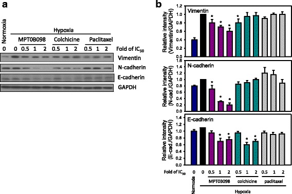
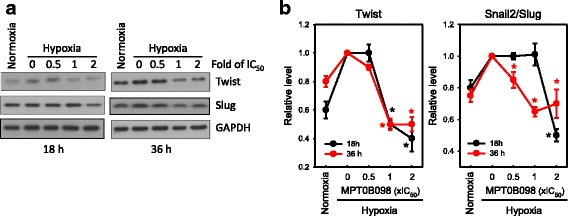
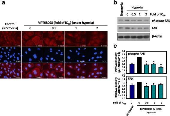
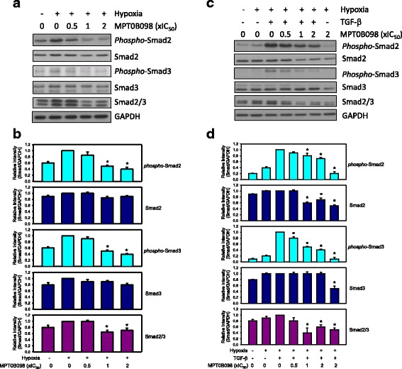
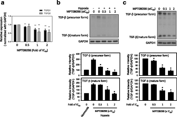
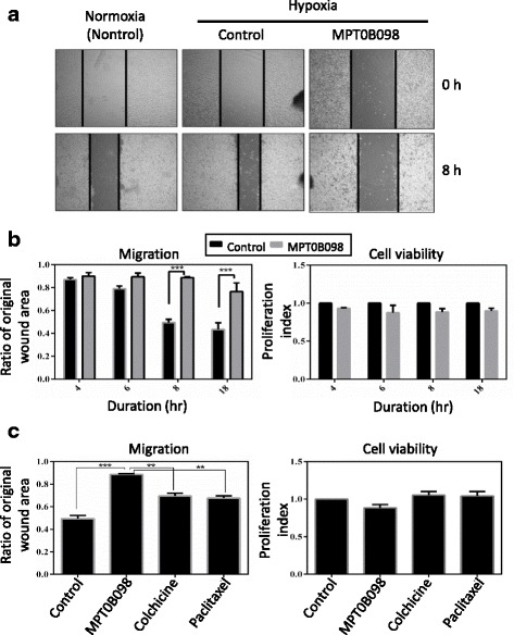
Results: MPT0B098 significantly inhibited HIF-1α expression, epithelial-to-mesenchymal morphology changes, and migratory ability in the human head and neck squamous cell carcinoma cell line OEC-M1. Furthermore, after MPT0B098 treatment, the expression of two mesenchymal markers, vimentin and N-cadherin, was downregulated under hypoxic conditions. Moreover, MPT0B098 suppressed hypoxia-induced EMT in part by inhibiting EMT-activating transcription factors, Twist and SNAI2/Slug. In addition, the inhibition of hypoxia-induced F-actin rearrangement and focal adhesion kinase phosphorylation may have contributed to suppression of EMT by MPT0B098in OEC-M1 cells. MPT0B098 significantly inhibited transforming growth factor(TGF)-β-induced phosphorylation of receptor-associated Smad2/3 by downregulating TGF-β mRNA and protein expression.

Conclusions: Taken together, this study provides a novel insight into the role of MPT0B098 in inhibiting hypoxia-induced EMT, suggesting its potential use for treating head and neck cancers.

Keywords: Epithelial to mesenchymal transition; Head and neck cancer; Hypoxia; Microtubule inhibitor; TGF-β.


Conflict of interest statement

Ethics approval and consent to participate

No applicable

Consent for publication

No applicable

Competing interests

The authors declare that they have no competing interests.

Publisher’s Note

Springer Nature remains neutral with regard to jurisdictional claims in published maps and institutional affiliations.

Figures

Fig. 1
Fig. 1
Antiproliferative effect of MPT0B098 in OEC-M1 cells under normoxic and hypoxic conditions. a The chemical structure of MPT0B098. b The in vitro antiproliferative activity of MPT0B098 in OEC-M1 cells under normoxic and hypoxic conditions. OEC-M1 cells were treated with MPT0B098, colchicine, or paclitaxel under normoxic and hypoxic conditions for 72 h. The IC50 values of these compounds resulting from 50% inhibition of cell growth were calculated using the methylene blue dye assay. Each value represents the mean ± SD of three independent experiments. c Hypoxia-induced drug resistance is the IC50 value of the test compounds in hypoxia divided by the equivalent in normoxia (* p < 0.05)
Fig. 2
Fig. 2
MPT0B098 inhibits hypoxia-induced EMT in OEC-M1 cells. a The effect of MPT0B098 onhypoxia-induced HIF-1αexpression. OEC-M1 cells were treated with various concentrations, indicated as fold of IC50 values, of MPT0B098 for 18 h under hypoxic conditions. At the end of the drug treatment, cell lysates were prepared and analyzed by SDS-PAGE and Western blot. β-Actin was used as an internal control. b Each bar depicts the mean of the relative intensity of HIF-1α from three independent experiments. c The effect of MPT0B098 on hypoxia-induced EMT.Cells were treated with MPT0B098 at a concentration of 0.5-fold IC50 for 48 h under hypoxic conditions and then cell morphology was examined by crystal violet staining. Cells in normoxia were used as controls
Fig. 3
Fig. 3
Effect of MPT0B098 and other microtubule inhibitors on the expression of EMT-related proteins in OEC-M1 cells under hypoxic conditions. a Parallel comparison of the effects of MPT0B098, colchicine, and paclitaxel on the expression of vimentin, N-cadherin, and E-cadherin in OEC-M1 cells.OEC-M1 cells were treated with various concentrations,indicated as fold of IC50 values,of MPT0B098, colchicine, or paclitaxel for 36 h under hypoxic conditions. At the end of the drug treatment, cell lysates were prepared and analyzed by SDS-PAGE and Western blot. GAPDH was used as an internal control. b Each bar depicts the mean of the relative intensity of vimentin, N-cadherin, and E-cadherin from three independent experiments (* p < 0.05, compared to hypoxia control).
Fig. 4
Fig. 4
MPT0B098 suppresses hypoxia-induced EMT by inhibiting the expression of EMT-activating transcription factors, Twist and SNAI2/Slug. a The effects of MPT0B098 on the expression of Twist and SNAI2/Slug in OEC-M1 cells.OEC-M1 cells were treated with various concentrations, indicated as fold of IC50 values, of MPT0B098 for 18 and 36 h under hypoxic conditions. At the end of the drug treatment, cell lysates were prepared and analyzed by SDS-PAGE and Western blot. GAPDH was used as an internal control. Each values depicts the mean of the relative intensities of Twist and SNAI2/Slug from three independent experiments. b Each value depicts the mean of the relative intensities of Twist and SNAI2/Slug from three independent experiments (* p < 0.05, compared to hypoxia control)
Fig. 5
Fig. 5
MPT0B098 inhibits hypoxia-induced F-actin rearrangement and FAK phosphorylation. a Immunofluorescent analysis of F-actin. OEC-M1 cells were treated with various concentrations, indicated as fold of IC50, of MPT0B098 for 18 h under hypoxic conditions, stained with phalloidin to label F-actin (red), counterstained with DAPI (blue), and then observed using an OLYMPUS florescence microscope. b Effect of MPT0B098 on FAK phosphorylation and expression. OEC-M1 cells were treated with various concentrations of MPT0B098 for 18 h.At the end of the drug treatment, cell lysates were prepared and analyzed by SDS-PAGE and Western blot. β-Actin was used as an internal control. c Each bar depicts the mean of the relative intensities of Twist and SNAI2/Slug from three independent experiments (* p < 0.05, compared to hypoxia control)
Fig. 6
Fig. 6
MPT0B098 downregulatesTGF-β/Smad signalingin OEC-M1 cells. a Cells were treated with various concentrations,indicated as fold of IC50 values, of MPT0B098 for 36 h under hypoxic conditions. b Each bar depicts the mean of the relative intensities of phospho-Smads and Smads from three independent experiments (* p < 0.05, compared to hypoxia control). c Cells were treated with various concentrations,indicated as fold of IC50 values, of MPT0B098 for 18 h, followed by an addition of 5 ng/mL TGF-β for another 18 h under hypoxic conditions. At the end of the drug treatment, cell lysates were prepared and analyzed by SDS-PAGE and Western blot. GAPDH was used as an internal control. d Each bar depicts the mean of the relative intensities of phospho-Smads and Smads from three independent experiments (* p < 0.05, compared to hypoxia control)
Fig. 7
Fig. 7
MPT0B098 downregulates TGF-β signaling by decreasing the expression of TGF-β mRNA and protein in OEC-M1 cells under hypoxic conditions. a Effect of MPT0B098 on the expression levels of TGF-β1 and TGF-β2 mRNA. Cells were treated with various concentrations of MPT0B098 in hypoxia. After incubation for 36 h, total RNA was extracted, reverse transcribed into cDNA, and subjected to PCR for detection of TGF-β1 and TGF-β2. GAPDH was used as an internal control. Data are represented as mean ± SD in triplicate. **P < 0.01 and ***P < 0.001for comparison between the control and treatment groups using an unpaired two-tailed Student’s t test. b Effect of MPT0B098 on the expression level of TGF-β protein. Cells were treated with various concentrations of MPT0B098 for 36 h in hypoxia. Quantification of TGF-β protein was determined by normalization with GAPDH (* p < 0.05, compared to hypoxia control). c The inhibitory effect of MPT0B098 on TGF-β under normoxic conditions. Cells were treated with MPT0B098 in normoxia at the indicated concentrations for 36 h. Quantification of TGF-β protein was determined by normalization with GAPDH (* p < 0.05, compared to control)
Fig. 8
Fig. 8
MPT0B098 inhibits hypoxia-induced cell migration in OEC-M1 cells. a Effect of MPT0B098 on cell motility. OEC-M1 cells were treated with 0.5-fold IC50 of MPT0B098 for 8 h and then cell motility was determined by the wound healing assay. b Quantification of cell migration was carried out by measuring the wound area (left panel) and calculating cell viability (right panel) after cells were treated with 0.5-fold IC50 of MPT0B098 for 4, 6, 8, and18 h. c Parallel comparison of the effect of MPT0B098 with other microtubule inhibitors, including colchicine and paclitaxel, at the drug concentration of 0.5-fold IC50on cell motility (left panel) and viability (right panel) at a treatment duration of 8 h. Data are represented as mean ± SD in triplicate. **P < 0.01 and ***P < 0.001 for comparison between the control and treatment groups using an unpaired two-tailed Student’s t test
Fig. 9
Fig. 9
Proposed pathway of MPT0B098-mediated EMT suppression. Blue solid line: proposed working models; orange dotted line: literature reported

Similar articles

Cited by

References

    1. Brown JM, Giaccia AJ. The unique physiology of solid tumors: opportunities (and problems) for cancer therapy. Cancer Res. 1998;58(7):1408–1416. - PubMed
    1. Bennewith KL, Dedhar S. Targeting hypoxic tumour cells to overcome metastasis. BMC Cancer. 2011;11:504. doi: 10.1186/1471-2407-11-504. - DOI - PMC - PubMed
    1. Ruas JL, Poellinger L. Hypoxia-dependent activation of HIF into a transcriptional regulator. Semin Cell Dev Biol. 2005;16(4–5):514–522. doi: 10.1016/j.semcdb.2005.04.001. - DOI - PubMed
    1. Thiery JP. Epithelial-mesenchymal transitions in tumour progression. Nat Rev Cancer. 2002;2(6):442–454. doi: 10.1038/nrc822. - DOI - PubMed
    1. Guan X. Cancer metastases: challenges and opportunities. Acta Pharm Sin B. 2015;5(5):402–418. doi: 10.1016/j.apsb.2015.07.005. - DOI - PMC - PubMed

MeSH terms