. 2018 Mar 28;15(1):98.
doi: 10.1186/s12974-018-1134-4.

The Aβ protofibril selective antibody mAb158 prevents accumulation of Aβ in astrocytes and rescues neurons from Aβ-induced cell death

Affiliations

The Aβ protofibril selective antibody mAb158 prevents accumulation of Aβ in astrocytes and rescues neurons from Aβ-induced cell death

Sofia Söllvander et al. J Neuroinflammation. .

Abstract

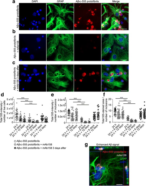
Background: Currently, several amyloid beta (Aβ) antibodies, including the protofibril selective antibody BAN2401, are in clinical trials. The murine version of BAN2401, mAb158, has previously been shown to lower the levels of pathogenic Aβ and prevent Aβ deposition in animal models of Alzheimer's disease (AD). However, the cellular mechanisms of the antibody's action remain unknown. We have recently shown that astrocytes effectively engulf Aβ42 protofibrils, but store rather than degrade the ingested Aβ aggregates. In a co-culture set-up, the incomplete degradation of Aβ42 protofibrils by astrocytes results in increased neuronal cell death, due to the release of extracellular vesicles, containing N-truncated, neurotoxic Aβ.

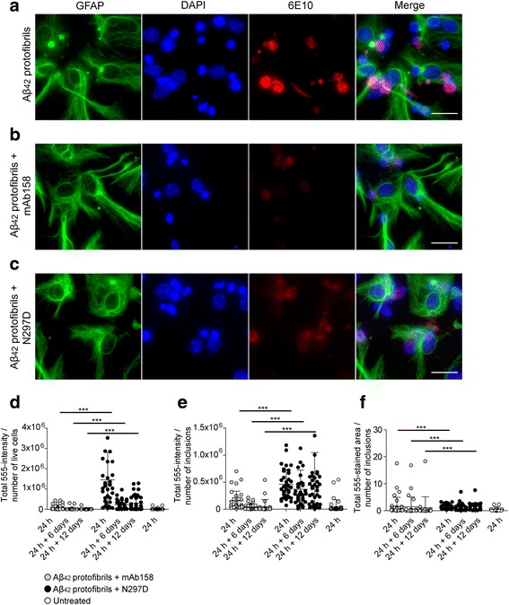
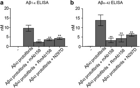
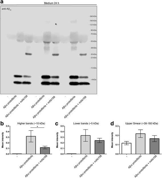
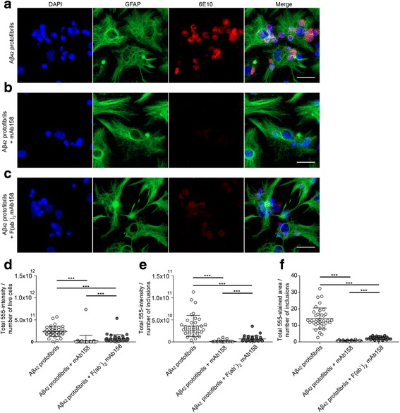
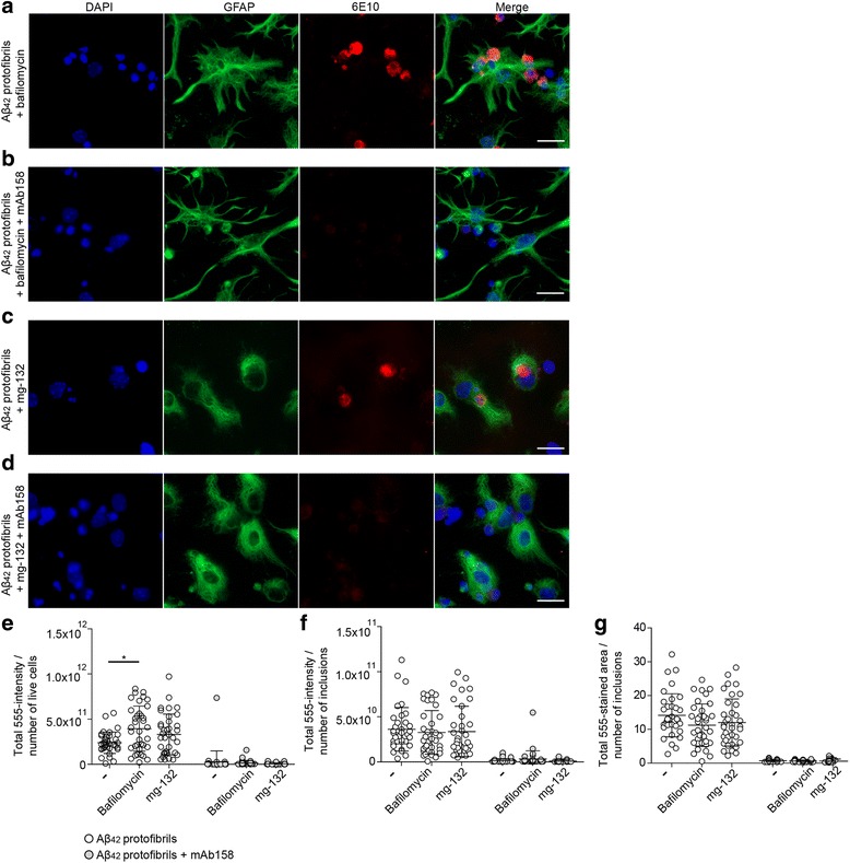
Methods: The aim of the present study was to investigate if the accumulation of Aβ in astrocytes can be affected by the Aβ protofibril selective antibody mAb158. Co-cultures of astrocytes, neurons, and oligodendrocytes, derived from embryonic mouse cortex, were exposed to Aβ42 protofibrils in the presence or absence of mAb158.

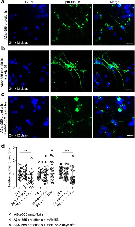
Results: Our results demonstrate that the presence of mAb158 almost abolished Aβ accumulation in astrocytes. Consequently, mAb158 treatment rescued neurons from Aβ-induced cell death.

Conclusion: Based on these findings, we conclude that astrocytes may play a central mechanistic role in anti-Aβ immunotherapy.

Keywords: Alzheimer’s disease; Amyloid-β; Antibody; Astrocyte; Clearance; Neuron.


Conflict of interest statement

Ethics approval and consent to participate

All experiments involving animals were performed at Uppsala University, Sweden. The experiments were approved by the Uppsala County Animal Ethics Board (ethical permit number: C75/13, valid 2013-06-28 to 2018-06-28), following the rules and regulations of the Swedish Animal Welfare Agency, in compliance with the European Communities Council Directive of 22 September 2010 (2010/63/EU).

Consent for publication

Not applicable.

Competing interests

LS is employed by BioArctic AB. LL is a co-founder of BioArctic AB and stock owner. This does not alter to the Journal of Neuroinflammation policies on sharing data and materials. None of the authors have any financial relationship with the organizations that sponsored the research. The other authors declare that they have no competing interests.

Publisher’s Note

Springer Nature remains neutral with regard to jurisdictional claims in published maps and institutional affiliations.

Figures

Fig. 1
Fig. 1
mAb158 reduces Aβ accumulation in astrocytes. Exposure of co-cultures to Aβ42-555 protofibrils for 24 h results in large Aβ inclusions in astrocytes (a). Concurrent addition of mAb158 clearly reduces the intracellular Aβ42-555 in astrocytes (b). However, mAb158 added 3 days after the Aβ42-555 protofibril exposure had no effect on Aβ42-555 accumulation (c). Using the Zen software, the 555-intensity and 555-stained area of the inclusions were measured. The 555-intensity/number of living cells (d), 555-intensity/number of Aβ inclusions (e), and total 555-stained area of Aβ-555 inclusions (f) decreased significantly in co-cultures treated with Aβ42-555 protofibrils + mAb158, compared to cultures exposed to Aβ42-555 protofibrils only or cultures treated with mAb158 3 days after Aβ42-555 protofibril exposure. Confocal imaging demonstrates intracellular co-localization of Aβ42-555 protofibrils and mAb158 in astrocyte (g). GFAP (green), DAPI (blue), mAb158 (white), and Aβ42-555 (red). The image is an orthogonal view of a confocal z-stack image. The XZ plane and YZ plane are shown at the top and to the right of the XY image, respectively. Scale bar 20 μm. The experiments were performed in triplicates with independent cell cultures, and 10 images/experiment were analyzed. Statistical analysis using Mann–Whitney U test (***P < 0.001)
Fig. 2
Fig. 2
mAb158 rescues neurons from Aβ-induced toxicity. Aβ42-555 protofibril exposure of co-cultures results in neuronal cell death 12 days following the removal of the Aβ42-555 protofibrils (a). Simultaneous addition of mAb158, significantly rescued the neurons (b), but this effect was not seen when the mAb158 was added 3 days after the Aβ42-555 protofibrils (c). The relative number of living neurons was significantly decreased 12 days following Aβ42-555 protofibril exposure (24 h + 12 days, **P < 0.01), but no neurotoxic effect was observed at 24 h + 12 days in cultures exposed to Aβ42-555 protofibrils together with mAb158. When mAb158 were added 3 days after the Aβ42-555 protofibril removal, no protective effect of mAb158 was found (***P < 0.001) (d). βIII-tubulin (green) and DAPI (blue). Scale bar 20 μm. The experiments were performed in triplicates with independent cell cultures, and 10 images/experiment were analyzed using Mann–Whitney U test
Fig. 3
Fig. 3
mAb158 reduces intracellular Aβ load, at least partly through an Fcγ receptor-independent pathway. Aβ42 protofibril accumulation in astrocytes (a) is reduced by preincubating Aβ42 protofibrils with mAb158 (b) or with N297D (c). Although, there was a clear reduction in Aβ accumulation with the presence of N297D as compared to Aβ42 protofibril exposed co-cultures, the cultures treated with mAb158 had significantly lower 555-intensity/number of living cells (d), 555-intensity/number of Aβ inclusions (e), and total 555-stained area of Aβ inclusions (f), compared to cultures treated with N297D. GFAP (green), DAPI (blue), and Aβ (red). Scale bar 20 μm. The experiments were performed in triplicates with independent cell cultures, and 10 images/experiment were analyzed. Statistical analysis using Mann–Whitney U test (***P < 0.001)
Fig. 4
Fig. 4
Anti-Aβ protofibril selective antibodies reduce the levels of intracellular Aβ. The Aβ levels in total cell lysates were measured using Aβ1-x and Aβx-42 ELISA. Both Aβ1-x (a) and Aβx-42 (b) levels were reduced in cultures treated with Aβ42 protofibrils + mAb158, Aβ42 protofibrils + RmAb158, or Aβ42 protofibrils + N297D, compared to cultures exposed to Aβ42 protofibrils only. All concentrations are expressed in nanomolar (nM) units. Mean values are from duplicates of three independent experiments, from two repeated analyses (**P < 0.01)
Fig. 5
Fig. 5
In the presence of mAb158, Aβ is cleared from the cell culture media. Western blot analysis showed a reduction of Aβ in the cell culture media of cultures exposed to Aβ42 protofibrils + mAb158, compared to cultures exposed to Aβ42 protofibril only (a). The reduction was seen both in low-molecular Aβ at ~ 10 kDa (b) and ~ 5 kDa (c) and in the smear of Aβ detected at ~ 38–160 kDa (d). Intensity measurements were performed from three independent cell culture experiments, and statistical analysis was performed using one-way ANOVA followed by Tukey’s multiple comparison test (*P < 0.05)
Fig. 6
Fig. 6
mAb158 F(ab’)2 fragments reduce Aβ accumulation in astrocytes. To confirm that the effect of mAb158 on Aβ42 protofibril degradation was a Fcγ receptor-independent mechanism, cell cultures were exposed to the F(ab’)2 fragments of mAb158 together with the Aβ42 protofibrils. Astrocytes in co-cultures exposed to Aβ42 protofibrils accumulated Aβ (a) but not when co-treated with mAb158 (b). In cultures treated with F(ab’)2 mAb158 + Aβ42 protofibrils, the accumulation of Aβ was reduced, almost to the same level as for mAb158 (c). The 555-intensity/number of living cells (d), 555-intensity/number of Aβ inclusions (e), and total 555-stained area of Aβ inclusions (f) were all significantly reduced in co-cultures treated with Aβ42 protofibril + mAb158 and Aβ42 protofibril + mAb158 F(ab’)2 fragments, as compared to Aβ42 protofibril exposed co-cultures. GFAP (green), DAPI (blue), and Aβ (red). Scale bar 20 μm. The experiments were performed in triplicates with independent cell cultures, and 10 images/experiment were analyzed using Mann–Whitney U test (***P < 0.001)
Fig. 7
Fig. 7
Proteosomal or endosomal-lysosomal inhibitors do not alter the mAb158-mediated effects on Aβ accumulation in astrocytes. Co-cultures were incubated with the proteosomal inhibitor mg-132 or the lysosomal inhibitor bafilomycin for 30 min prior to the Aβ42 protofibril exposure and mAb158 treatment. The inhibitors were also present during the 24-h exposure. Neither of the inhibitors affected the mAb158-mediated reduction of Aβ deposits (ad). Measurement of 555-intensity/number of living cells (e), 555-intensity/number of Aβ inclusions (f), and total 555-stained area of Aβ inclusions (g) demonstrated that there was a significant reduction in Aβ inclusions in Aβ42 protofibril-exposed cultures when co-treated with mAb158 + bafilomycin or mAb158 + mg-132. This confirms that the inhibitors did not interfere with the action of mAb158. GFAP (green), DAPI (blue), and Aβ (red). Scale bar 20 μm. The experiments were performed in triplicates with independent cell cultures, and 10 images/experiment were analyzed using Mann–Whitney U test (**P < 0.01)

Similar articles

Cited by

References

    1. Walsh DM, Klyubin I, Fadeeva JV, Cullen WK, Anwyl R, Wolfe MS, Rowan MJ, Selkoe DJ. Naturally secreted oligomers of amyloid beta protein potently inhibit hippocampal long-term potentiation in vivo. Nature. 2002;416:535–539. doi: 10.1038/416535a. - DOI - PubMed
    1. Lee SJ, Nam E, Lee HJ, Savelieff MG, Lim MH. Towards an understanding of amyloid-beta oligomers: characterization, toxicity mechanisms, and inhibitors. Chem Soc Rev. 2017;46:310–323. doi: 10.1039/C6CS00731G. - DOI - PubMed
    1. Williams TL, Serpell LC. Membrane and surface interactions of Alzheimer's Abeta peptide—insights into the mechanism of cytotoxicity. FEBS J. 2011;278:3905–3917. doi: 10.1111/j.1742-4658.2011.08228.x. - DOI - PubMed
    1. Klyubin I, Walsh DM, Cullen WK, Fadeeva JV, Anwyl R, Selkoe DJ, Rowan MJ. Soluble Arctic amyloid beta protein inhibits hippocampal long-term potentiation in vivo. Eur J Neurosci. 2004;19:2839–2846. doi: 10.1111/j.1460-9568.2004.03389.x. - DOI - PubMed
    1. Hartley DM, Walsh DM, Ye CP, Diehl T, Vasquez S, Vassilev PM, Teplow DB, Selkoe DJ. Protofibrillar intermediates of amyloid beta-protein induce acute electrophysiological changes and progressive neurotoxicity in cortical neurons. J Neurosci. 1999;19:8876–8884. - PMC - PubMed

Substances