Immuno-modulatory effect of local rhEGF treatment during tissue repair in diabetic ulcers
- PMID: 29592858
- PMCID: PMC5900456
- DOI: 10.1530/EC-18-0117
Immuno-modulatory effect of local rhEGF treatment during tissue repair in diabetic ulcers
Abstract
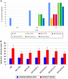
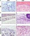
Wound healing is a complex process that can be severely impaired due to pathological situations such as diabetes mellitus. Diabetic foot ulcers are a common complication of this pathology and are characterized by an excessive inflammatory response. In this work, the effects of local treatment with recombinant human epidermal growth factor (rhEGF) were studied using a full-thickness wound healing model in streptozotocin-induced diabetic rats. Wound healing process was assessed with different concentrations of rhEGF (0.1, 0.5, 2.0 and 8.0 µg/mL), placebo and both diabetic and non-diabetic controls (n = 53). The macroscopic healing observed in treated diabetic rats was affected by rhEGF concentration. Histologically, we also observed an improvement in the epithelialization, granulation tissue formation and maturation in treated groups, finding again the best response at doses of 0.5 and 2.0 µg/mL. Afterwards, the tissue immune response over time was assessed in diabetic rats using the most effective concentrations of rhEGF (0.5 and 2.0 µg/mL), compared to controls. The presence of macrophages, CD4+ T lymphocytes and CD8+ T lymphocytes, in the reparative tissue was quantified, and cytokine expression was measured by quantitative real-time PCR. rhEGF treatment caused a reduction in the number of infiltrating macrophages in the healing tissue of diabetic, as well as diminished activation of these leukocytes. These findings show that local administration of rhEGF improves the healing process of excisional wounds and the quality of the neoformed tissue in a dose-dependent manner. Besides, this treatment reduces the local inflammation associated with diabetic healing, indicating immuno-modulatory properties.
Keywords: diabetes mellitus; diabetic foot ulcer; rhEGF; wound healing.
© 2018 The authors.
Figures


Similar articles
-
The effect of dextrin-rhEGF on the healing of full-thickness, excisional wounds in the (db/db) diabetic mouse.J Control Release. 2011 Jun 30;152(3):411-7. doi: 10.1016/j.jconrel.2011.03.016. Epub 2011 Mar 22. J Control Release. 2011. PMID: 21435363
-
Efficacy of intralesional recombinant human epidermal growth factor in diabetic foot ulcers in Mexican patients: a randomized double-blinded controlled trial.Wound Repair Regen. 2014 Jul-Aug;22(4):497-503. doi: 10.1111/wrr.12187. Wound Repair Regen. 2014. PMID: 25041620 Clinical Trial.
-
Nanotechnology promotes the full-thickness diabetic wound healing effect of recombinant human epidermal growth factor in diabetic rats.Wound Repair Regen. 2010 Sep-Oct;18(5):499-505. doi: 10.1111/j.1524-475X.2010.00612.x. Wound Repair Regen. 2010. PMID: 20840519
-
Efficacy of Topical Recombinant Human Epidermal Growth Factor for Treatment of Diabetic Foot Ulcer: A Systematic Review and Meta-Analysis.Int J Low Extrem Wounds. 2016 Jun;15(2):120-5. doi: 10.1177/1534734616645444. Epub 2016 May 5. Int J Low Extrem Wounds. 2016. PMID: 27151755
-
Topical Recombinant Human Epidermal Growth Factor for Diabetic Foot Ulcers: A Meta-Analysis of Randomized Controlled Clinical Trials.Ann Vasc Surg. 2020 Jan;62:442-451. doi: 10.1016/j.avsg.2019.05.041. Epub 2019 Aug 5. Ann Vasc Surg. 2020. PMID: 31394225
Cited by
-
Zuyangping formula promotes skin wound healing in diabetic rats.J Tradit Chin Med. 2024 Dec;44(6):1194-1203. doi: 10.19852/j.cnki.jtcm.2024.06.008. J Tradit Chin Med. 2024. PMID: 39617705 Free PMC article.
-
The efficacy of skin soft tissue expansion and recombinant human epidermal growth factor in the repair of second-degree scald scars: a prospective single-blind randomized controlled trial.Ann Surg Treat Res. 2025 May;108(5):325-330. doi: 10.4174/astr.2025.108.5.325. Epub 2025 Apr 28. Ann Surg Treat Res. 2025. PMID: 40352800 Free PMC article.
-
EGF, a veteran of wound healing: highlights on its mode of action, clinical applications with focus on wound treatment, and recent drug delivery strategies.Arch Pharm Res. 2023 Apr;46(4):299-322. doi: 10.1007/s12272-023-01444-3. Epub 2023 Mar 16. Arch Pharm Res. 2023. PMID: 36928481 Review.
-
Single-cell RNA-seq in diabetic foot ulcer wound healing.Wound Repair Regen. 2024 Nov-Dec;32(6):880-889. doi: 10.1111/wrr.13218. Epub 2024 Sep 12. Wound Repair Regen. 2024. PMID: 39264020 Free PMC article. Review.
References
-
- Fazeli Farsani S, van der Aa MP, van der Vorst MMJ, Knibbe CAJ, de Boer A. Global trends in the incidence and prevalence of type 2 diabetes in children and adolescents: a systematic review and evaluation of methodological approaches. Diabetologia 2013. 56 1471–1488. (10.1007/s00125-013-2915-z) - DOI - PubMed
-
- Centers for Disease Control and Prevention. Diabetes 2014 Report Card. Atlanta, GA, USA: CDC, 2015. (available at: https://www.cdc.gov/diabetes/pdfs/library/diabetesreportcard2014.pdf)
LinkOut - more resources
Full Text Sources
Other Literature Sources
Research Materials

