. 2018 Jun;24(6):828-840.
doi: 10.1261/rna.064659.117. Epub 2018 Mar 28.

Decreased A-to-I RNA editing as a source of keratinocytes' dsRNA in psoriasis

Affiliations

Decreased A-to-I RNA editing as a source of keratinocytes' dsRNA in psoriasis

Lea Shallev et al. RNA. 2018 Jun.

Abstract

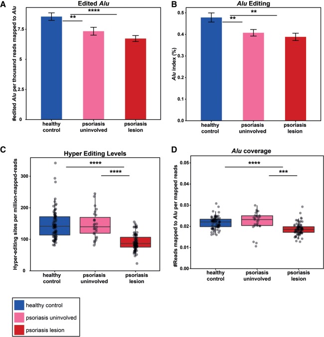
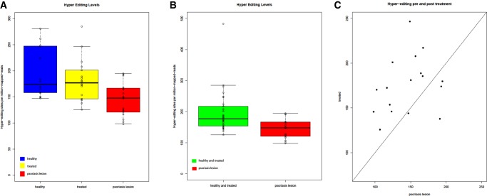
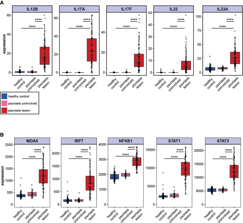
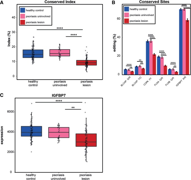
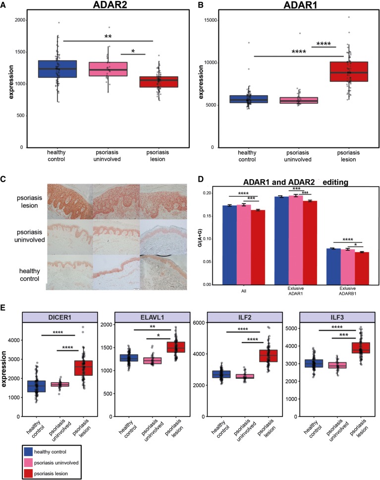
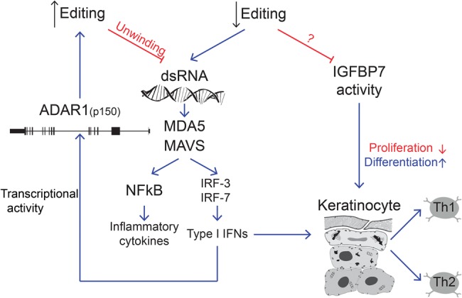
Recognition of dsRNA molecules activates the MDA5-MAVS pathway and plays a critical role in stimulating type-I interferon responses in psoriasis. However, the source of the dsRNA accumulation in psoriatic keratinocytes remains largely unknown. A-to-I RNA editing is a common co- or post-transcriptional modification that diversifies adenosine in dsRNA, and leads to unwinding of dsRNA structures. Thus, impaired RNA editing activity can result in an increased load of endogenous dsRNAs. Here we provide a transcriptome-wide analysis of RNA editing across dozens of psoriasis patients, and we demonstrate a global editing reduction in psoriatic lesions. In addition to the global alteration, we also detect editing changes in functional recoding sites located in the IGFBP7, COPA, and FLNA genes. Accretion of dsRNA activates autoimmune responses, and therefore the results presented here, linking for the first time an autoimmune disease to reduction in global editing level, are relevant to a wide range of autoimmune diseases.

Keywords: A-to-I; RNA editing; interferon; psoriasis.


Figures

FIGURE 1.
FIGURE 1.
A decrease in global editing in psoriatic lesions. (A) The relative number of edited Alu in healthy controls (blue) is higher than in psoriatic lesion (red) and uninvolved skin (pink) samples. The bar-plot represents the average ±SEM of the number of edited Alu per thousand reads that were aligned to Alu. P-value (healthy versus psoriasis) = 9.2 × 10⁻⁶. (B) Measuring the Alu editing index in psoriatic lesions, uninvolved skin and healthy control tissues emphasizes the decreased editing activity in psoriasis. P-value = 0.001 (C) Complementary hyperediting screening reveals a decrease in the number of editing sites in psoriatic lesions. The distribution of the normalized number of hyperediting sites is shown. P-value = 2.6 × 10⁻¹⁷. (D) The relative expression of Alu repeats is lower in psoriatic tissues compared to normal controls, suggesting that the source for an elevated level of dsRNA is not an increased expression of dsRNA-forming transcripts. P-value = 7×10⁻¹¹; all analyses are two-tailed t-tests.
FIGURE 2.
FIGURE 2.
A second, independent set of psoriatic RNA-seq data show a decrease of editing in psoriatic lesions. (A) Total RNA-seq data set analysis repeats the trend of a decrease in the number of hyperediting sites in psoriatic lesions. Interestingly, after 1 mo of treatment with adalimumab, the editing levels restored back to a similar level of healthy control samples. The figure shows the distribution of the normalized number of hyperediting sites. P-value = 0.008, two-tailed t-test. (B) Healthy control and treated with adalimumab after 1-mo groups were joined together in order to achieve a better statistical power. Complementary hyperediting screening reveals a decrease in the number of editing sites in psoriatic lesions. P-value = 0.0004, two-tailed t-test. (C) Matched comparison of hyperediting in a sample between before and after treatment. P-value = 0.004, two-tailed t-test.
FIGURE 3.
FIGURE 3.
Overexpression of MDA5 pathway and IFN stimulated gene signature in psoriatic lesions. (A) Up-regulation of known Th1 and Th17 cytokines signature in psoriasis. Distribution of IL12B, IL17A, IL17F, IL22, and IL23A gene expression in psoriatic lesions (red), uninvolved skin (pink), and healthy control (blue) samples. Expression levels are represented by the normalized values from DESeq analysis. (B) Elevated mRNA expression of genes in the MDA5 pathway: MDA5 (P-value = 7.7 × 10⁻²⁰ and 4.0 × 10−57, respectively, two tailed t-test); IRF7 (P-value = 1.5 × 10⁻¹⁷ and 2.6 × 10−44, respectively, two tailed t-test); NFKB1 (P-value = 5.8 × 10⁻⁶ and 5.4 × 10⁻¹⁶, respectively, two tailed t-test); STAT1 (P-value = 9.9 × 10−50 and 5.0 × 10−136, respectively, two tailed t-test); and STAT3 (P-value = 5.2 × 10⁻²⁴ and 5 × 10−56, respectively, two tailed t-test).
FIGURE 4.
FIGURE 4.
Editing of conserved sites is reduced in psoriatic lesions. (A) The editing index of the conserved sites displays a lower signal in psoriatic lesions compared to uninvolved skin and healthy control samples. (B) Significantly decreased editing in conserved sites in psoriatic lesions. The editing activity in the sites that exhibit higher editing leads to nonsynonymous mutations in the COPA, FLNA, and IGFBP7 proteins. (C) IGFBP7 expression distribution in psoriatic lesions, uninvolved skin and healthy control samples. Down-regulation of IGFBP7 in psoriatic lesions as previously reported in the literature emphasizes the significant involvement of the gene in the pathology of psoriasis.
FIGURE 5.
FIGURE 5.
Expression of ADARs and possible regulation. Based on the RNA-seq data, ADAR2 is decreased (A) while ADAR1 is increased (B) in psoriatic lesions. (C) Up-regulation of ADAR1 was also observed at the protein level based on histochemical staining. (D) Global editing levels based on a set of 6583 known editing sites, and subgroups of this set that were reported to be specifically edited by ADAR1 (n = 4458) and by ADAR2 (n = 502). There is a common trend of editing decrease in targets of both ADAR enzymes. (E) Up-regulation of ADAR1-binding proteins’ expression in psoriatic lesions may provide a possible explanation for the decrease in editing in psoriatic lesions, and the observation emphasizes the complexity of A-to-I RNA editing regulation. DICER1: P-value = 1.4 × 10⁻⁷ and 1.8 × 10⁻¹⁷, respectively. ELAVL1: P-value = 0.025 and 0.005, respectively. ILF2: P-value = 7.6 × 10⁻⁷ and 8.0 × 10⁻¹¹, respectively. ILF3: P-value = 0.0007 and 9.6 × 10⁻⁶, respectively. All analyses are two tailed t-tests.
FIGURE 6.
FIGURE 6.
Proposed model for the A-to-I RNA editing involvement in psoriasis. Decreased editing in coding regions modulates genes such as IGFBP7 and COPA. In addition, the reduction in global editing triggers the accumulation of dsRNA molecules that activate the MDA5/MAVS pathway and stimulate type I IFN responses as well as NFkB associated cytokine production. A feedback mechanism up-regulates ADAR1.

Similar articles

Cited by

References

    1. Afshar M, Martinez AD, Gallo RL, Hata TR. 2013. Induction and exacerbation of psoriasis with Interferon-α therapy for hepatitis C: a review and analysis of 36 cases. J Eur Acad Dermatol Venereol 27: 771–778. - PMC - PubMed
    1. Ahn R, Gupta R, Lai K, Chopra N, Arron ST, Liao W. 2016. Network analysis of psoriasis reveals biological pathways and roles for coding and long non-coding RNAs. BMC Genomics 17: 841. - PMC - PubMed
    1. Alwawi EA, Mehlis SL, Gordon KB. 2008. Treating psoriasis with adalimumab. Ther Clin Risk Manag 4: 345–351. - PMC - PubMed
    1. Anders S, Huber W. 2010. Differential expression analysis for sequence count data. Genome Biol 11: R106. - PMC - PubMed
    1. Andrews S. 2010. FastQC: A quality control tool for high throughput sequence data. Vol. 2011. http://www.bioinformatics.babraham.ac.uk/projects/fastqc

Publication types

MeSH terms