. 2018 Apr 10;2(7):731-744.
doi: 10.1182/bloodadvances.2017015602.

Selectins and chemokines use shared and distinct signals to activate β2 integrins in neutrophils

Affiliations

Selectins and chemokines use shared and distinct signals to activate β2 integrins in neutrophils

Tadayuki Yago et al. Blood Adv. .

Abstract

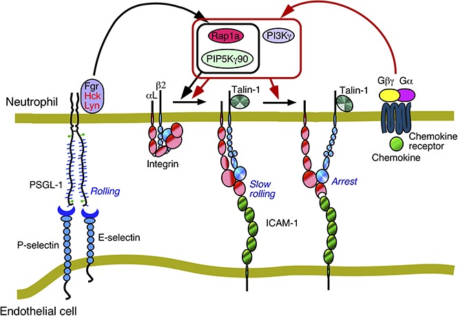
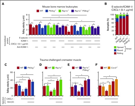
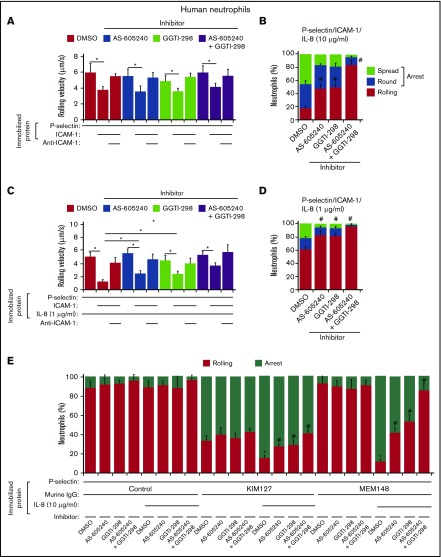
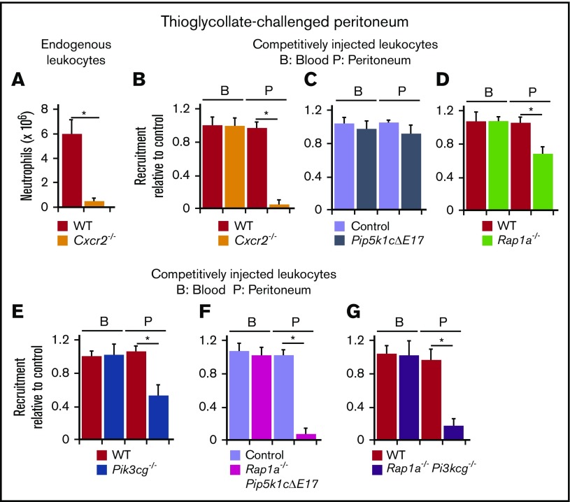
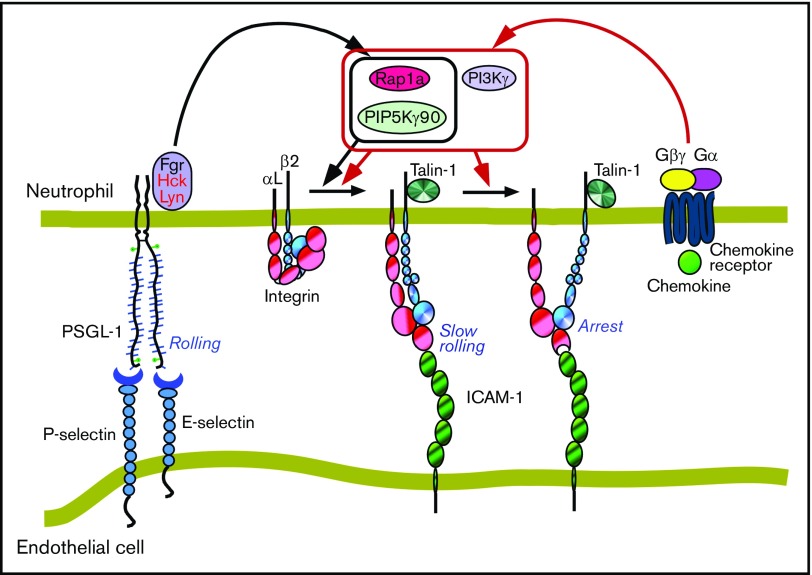
Rolling neutrophils receive signals while engaging P- and E-selectin and chemokines on inflamed endothelium. Selectin signaling activates β2 integrins to slow rolling velocities. Chemokine signaling activates β2 integrins to cause arrest. Despite extensive study, key aspects of these signaling cascades remain unresolved. Using complementary in vitro and in vivo assays, we found that selectin and chemokine signals in neutrophils triggered Rap1a-dependent and phosphatidylinositol-4-phosphate 5-kinase γ (PIP5Kγ90)-dependent pathways that induce integrin-dependent slow rolling and arrest. Interruption of both pathways, but not either pathway alone, blocked talin-1 recruitment to and activation of integrins. An isoform of PIP5Kγ90 lacking the talin-binding domain (PIP5Kγ87) could not activate integrins. Chemokines, but not selectins, used phosphatidylinositol-4,5-bisphosphate 3-kinase γ (PI3Kγ) in cooperation with Rap1a to mediate integrin-dependent slow rolling (at low chemokine concentrations), as well as arrest (at high chemokine concentrations). High levels of chemokines activated β2 integrins without selectin signals. When chemokines were limiting, they synergized with selectins to activate β2 integrins.


Conflict of interest statement

Conflict-of-interest disclosure: R.P.M. is a cofounder of Selexys Pharmaceuticals, now part of Novartis AG, and of Tetherex Pharmaceuticals. The remaining authors declare no competing financial interests.

Figures

None
Graphical abstract
Figure 1.
Figure 1.
Selectin-interacting mouse leukocytes from bone marrow or peripheral blood are mature neutrophils. (A-E) Scatter profiles of the indicated mouse leukocyte populations. A gate (red circle in each panel) was set around cells expressing high levels of Ly6G. (F-K) Expression levels of the indicated surface protein in the gated populations. The colored histograms numbered 1 to 5 in each panel correspond to the cells in A-E. (L) Rolling velocities of cells from the indicated population on E-selectin with or without co-immobilized ICAM-1 in the presence or absence of anti-ICAM-1 mAb, the Src family kinase inhibitor PP2, or its inactive analog PP3. (M) Percentages of cells from the indicated population rolling, arrested and round, or arrested and spread on co-immobilized E-selectin, ICAM-1, and CXCL1 in the presence of the chemokine receptor inhibitor PTx or its solvent control dimethyl sulfoxide (DMSO). The histograms in A-K are representative of 5 independent experiments. The data in L and M represent the mean ± SEM from 5 experiments, with 5 mice in each experimental group. *P < .05 for rolling velocity; #P < .05 for percentage of rolling cells compared with that in WT, as determined by unpaired Student t test.
Figure 2.
Figure 2.
Rap1a and PIP5Kγ90 cooperate to induce neutrophil slow rolling and arrest. (A,C,E,G,I) Rolling velocities of neutrophils of the indicated genotype on E-selectin with or without co-immobilized ICAM-1 in the presence or absence of anti-ICAM-1 mAb. (B,D,F,H,J) Percentages of neutrophils of the indicated genotype rolling, arrested and round, or arrested and spread on co-immobilized E-selectin, ICAM-1, and CXCL1. (K) Isolated bone marrow neutrophils of the indicated genotype were incubated with or without CXCL1, lysed, and immunoprecipitated (IP) with control or anti-β2 integrin mAb. Immunoprecipitates were analyzed by immunoblotting (IB) with anti-talin or anti-β2 integrin antibodies. (L-N) Numbers of differentially labeled adherent bone marrow leukocytes from the indicated genotype in TNF-stimulated venules of cremaster muscle. In some experiments, labeled leukocytes were pretreated with PTx and then injected into TNF-challenged mice that were previously injected with PTx. (O-Q) Velocities of differentially labeled bone marrow leukocytes from mice of the indicated genotype rolling in TNF-stimulated venules of cremaster muscle, measured before and after injecting a blocking mAb to P-selectin and then a blocking mAb to β2 integrins. The labeled leukocytes were pretreated with PTx and then injected into TNF-challenged WT mice that were previously injected with PTx. The data in K are representative of 3 experiments. Other data represent the mean ± SEM from 5 experiments, with 5 mice in each experimental group. *P < .05 for rolling velocity; #P < .05 for percentage of rolling cells compared with that in WT, as determined by unpaired Student t test.
Figure 3.
Figure 3.
Rap1a and PI3Kγ in neutrophils cooperate to mediate chemokine-triggered, but not selectin-triggered, slow rolling and chemokine-triggered arrest. (A,C) Rolling velocities of neutrophils of the indicated genotype on E-selectin or P-selectin with or without co-immobilized ICAM-1 in the presence or absence of anti-ICAM-1 mAb. (B,D) Percentages of neutrophils of the indicated genotype rolling, arrested and round, or arrested and spread on co-immobilized E-selectin or P-selectin, ICAM-1, and CXCL1. (E-G) Velocities of differentially labeled bone marrow leukocytes from mice of the indicated genotype rolling in TNF-stimulated venules of cremaster muscle, measured before and after injecting a blocking mAb to P-selectin and then a blocking mAb to β2 integrins. The labeled leukocytes were pretreated with PTx and then injected into TNF-challenged WT mice that were previously injected with PTx. (H-J) Numbers of differentially labeled adherent bone marrow leukocytes from the indicated genotype in TNF-stimulated venules of cremaster muscle. The labeled bone marrow leukocytes were injected into TNF-challenged WT mice. The data represent the mean ± SEM from 5 experiments, with 5 mice in each experimental group. *P < .05 for rolling velocity; #P < .05 for percentage of rolling cells compared with that in WT, as determined by unpaired Student t test.
Figure 4.
Figure 4.
Rap1a and PI3Kγ in neutrophils cooperate to mediate chemokine-triggered, but not selectin-triggered, slow rolling and chemokine-triggered arrest. (A) Rolling velocities of neutrophils of the indicated genotype on E-selectin co-immobilized with ICAM-1 and low-dose CXCL1 (0.1 μg/mL) in the presence or absence of anti-ICAM-1 mAb. (B) Percentages of neutrophils of the indicated genotype rolling, arrested and round, or arrested and spread on co-immobilized E-selectin, ICAM-1, and low-dose CXCL1. (C-F) Velocities of differentially labeled bone marrow leukocytes from mice of the indicated genotype rolling in trauma-challenged venules of cremaster muscle of WT mice, measured before and after injection of CXCL1 (50 ng) and then a blocking mAb to β2 integrins. The data represent the mean ± SEM from 5 experiments, with 5 mice in each experimental group. *P < .05 for rolling velocity; #P < .05 for percentage of rolling cells compared with that in WT, as determined by unpaired Student t test.
Figure 5.
Figure 5.
Chemokines, but not selectins, cooperatively use Rap1 and PI3Kγ to trigger β2 integrin activation. (A) Rolling velocities of human neutrophils treated with the Rap1 inhibitor GGTI-298 and/or the PI3Kγ inhibitor AS-605240 on P-selectin with or without co-immobilized ICAM-1 in the presence or absence of anti-ICAM-1 mAb. (B) Percentages of human neutrophils treated with the indicated inhibitor rolling, arrested and round, or arrested and spread on co-immobilized P-selectin, ICAM-1, and IL-8 (10 μg/mL). (C) Rolling velocities of human neutrophils treated with the indicated inhibitor on P-selectin with co-immobilized ICAM-1 and low-dose IL-8 (1 μg/mL) in the presence or absence of anti-ICAM-1 mAb. (D) Percentages of human neutrophils treated with the indicated inhibitor rolling, arrested and round, or arrested and spread on co-immobilized P-selectin, ICAM-1, and low dose IL-8 (1 μg/mL). (E) Percentages of human neutrophils treated with the indicated inhibitor rolling or arrested on P-selectin co-immobilized with control IgG, KIM127, or MEM148 with or without IL-8 (10 μg/mL). The data represent the mean ± SEM from 5 experiments. *P < .05 for rolling velocity; #P < .05 for percentage of rolling cells compared with that in WT, as determined by unpaired Student t test.
Figure 6.
Figure 6.
Neutrophils require chemokine but not selectin signaling to migrate into the peritoneum after thioglycollate challenge. (A) WT or Cxcr2−/− mice were injected intraperitoneally with thioglycollate. After 2 hours, peritoneal cells were collected and the number of neutrophils was measured by flow cytometry. Neutrophils were identified by their scatter properties and by staining with anti-Ly6G mAb. (B-G) WT mice were injected intraperitoneally with thioglycollate. After 2 hours, they were injected intravenously with a 1:1 mixture of PKH-67-labeled WT bone marrow leukocytes and PKH-26-labeled bone marrow leukocytes of the indicated genotype. After another 2 hours, blood and peritoneal cells were collected, and the number of neutrophils labeled with each dye was measured by flow cytometry. Neutrophils were identified by their scatter properties and by staining with anti-Ly6G mAb. Results are plotted as the ratio of PKH-26-labeled neutrophils from the indicated genotype to PKH-67-labeled WT neutrophils. The data represent the mean ± SEM from 5 to 8 mice in each experimental group. *P < .05, as determined by unpaired Student test.
Figure 7.
Figure 7.
Selectin and chemokine signaling pathways in neutrophils. Each arrow indicates a signaling outcome. Signaling intermediates are omitted for clarity. See “Discussion” for details.

Similar articles

Cited by

References

    1. Ley K, Laudanna C, Cybulsky MI, Nourshargh S. Getting to the site of inflammation: the leukocyte adhesion cascade updated. Nat Rev Immunol. 2007;7(9):678-689. - PubMed
    1. McEver RP, Zhu C. Rolling cell adhesion. Annu Rev Cell Dev Biol. 2010;26(1):363-396. - PMC - PubMed
    1. McEver RP. Selectins: initiators of leucocyte adhesion and signalling at the vascular wall. Cardiovasc Res. 2015;107(3):331-339. - PMC - PubMed
    1. Lefort CT, Ley K. Neutrophil arrest by LFA-1 activation. Front Immunol. 2012;3:157. - PMC - PubMed
    1. Lagarrigue F, Kim C, Ginsberg MH. The Rap1-RIAM-talin axis of integrin activation and blood cell function. Blood. 2016;128(4):479-487. - PMC - PubMed

Publication types

Substances