. 2018 Jul;16(7):1092-1102.
doi: 10.1158/1541-7786.MCR-18-0106. Epub 2018 Mar 28.

Synthetic Lethality of PARP Inhibition and Ionizing Radiation is p53-dependent

Affiliations

Synthetic Lethality of PARP Inhibition and Ionizing Radiation is p53-dependent

Steven T Sizemore et al. Mol Cancer Res. 2018 Jul.

Abstract

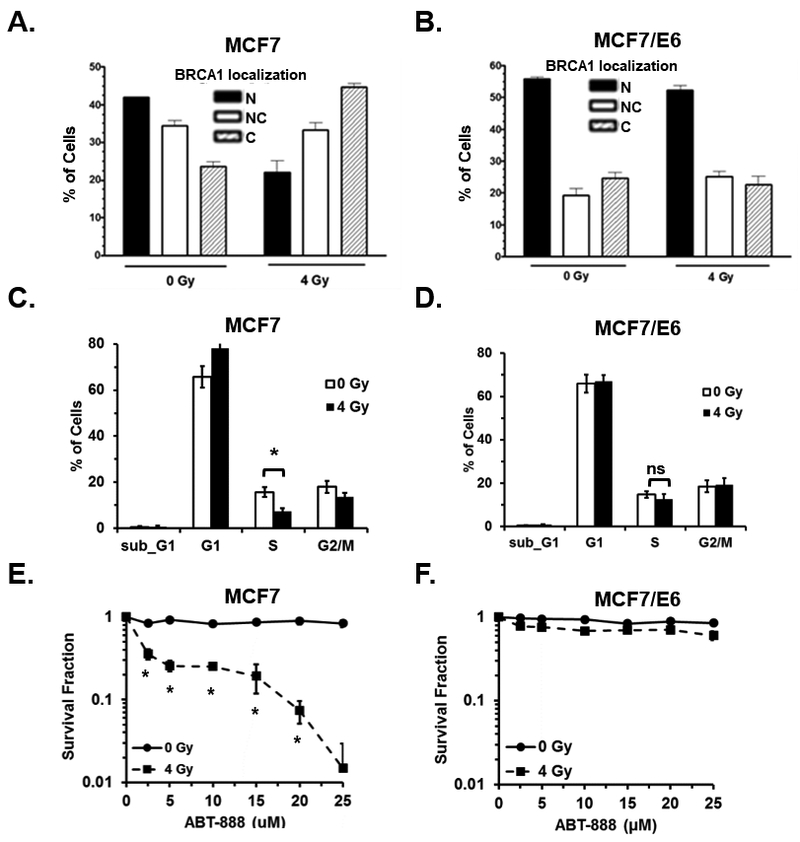
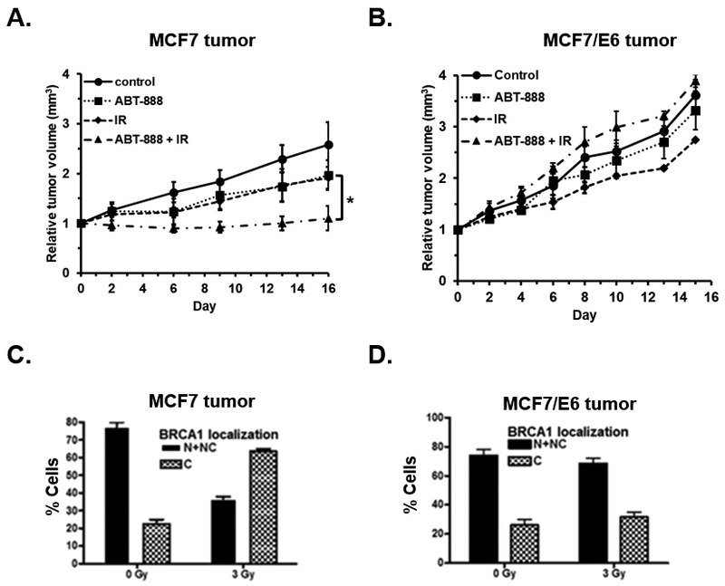
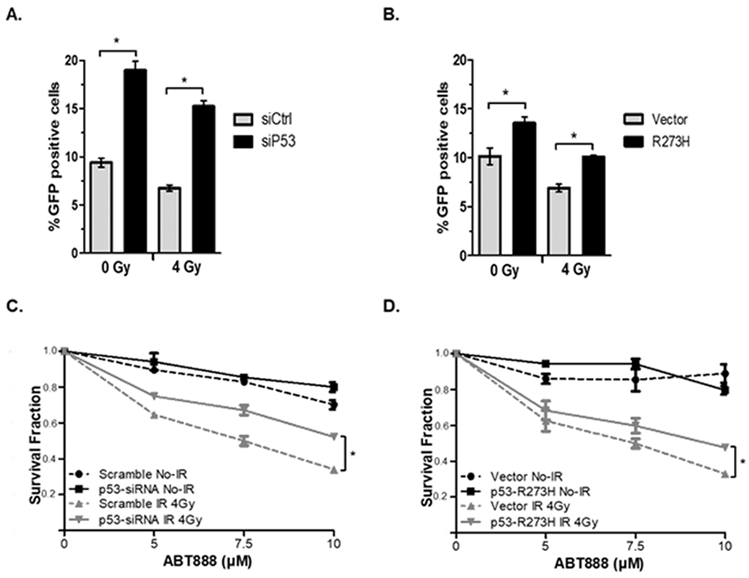
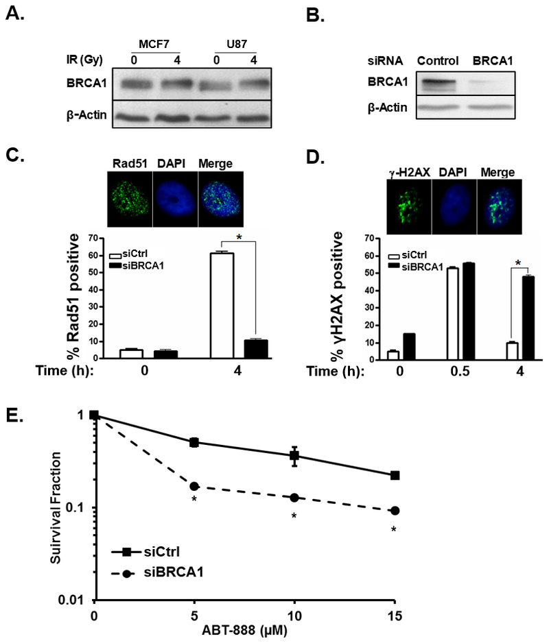
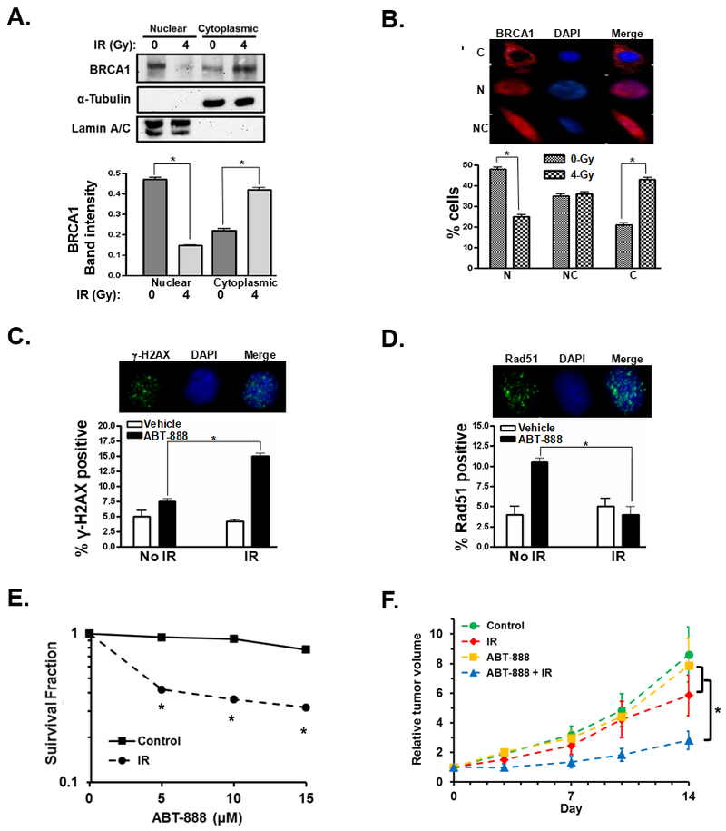
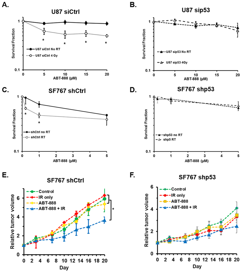
PARP inhibitors (PARPi) are potentially effective therapeutic agents capable of inducing synthetic lethality in tumors with deficiencies in homologous recombination (HR)-mediated DNA repair such as those carrying BRCA1 mutations. However, BRCA mutations are rare, the majority of tumors are proficient in HR repair, and thus most tumors are resistant to PARPi. Previously, we observed that ionizing radiation (IR) initiates cytoplasmic translocation of BRCA1 leading to suppression of HR-mediated DNA repair and induction of synthetic PARPi lethality in wild-type BRCA1 and HR-proficient tumor cells. The tumor suppressor p53 was identified as a key factor that regulates DNA damage-induced BRCA1 cytoplasmic sequestration following IR. However, the role of p53 in IR-induced PARPi sensitization remains unclear. This study elucidates the role of p53 in IR-induced PARPi cytotoxicity in HR-proficient cancer cells and suggests p53 status may help define a patient population that might benefit from this treatment strategy. Sensitization to PARPi following IR was determined in vitro and in vivo utilizing human breast and glioma tumor cells carrying wild-type BRCA1 and p53, and in associated cells in which p53 function was modified by knockdown or mutation. In breast and glioma cells with proficient HR repair, IR-induced BRCA1 cytoplasmic sequestration, HR repair inhibition, and subsequent PARPi sensitization in vitro and in vivo was dependent upon functional p53.Implications: Implications: p53 status determines PARP inhibitor sensitization by ionizing radiation in multiple BRCA1 and HR-proficient tumor types and may predict which patients are most likely to benefit from combination therapy. Mol Cancer Res; 16(7); 1092-102. ©2018 AACR.


Conflict of interest statement

Conflicts of Interest: No author claims a conflict of interest.

Figures

Figure 1.
Figure 1.. p53 function is required for IR-induced cytoplasmic translocation of BRCA1 and sensitization of breast cancer cells to PARPi in vitro.
A and B, the percentage of MCF7 control (A) and MCF7/E6 (B) cells that demonstrated nuclear (N), nuclear and cytoplasmic (NC), or cytoplasmic (C) immunohistochemical staining of BRCA1 was analyzed 24 h after treatment with 0 or 4 Gy IR. Results are the average of three independent experiments. C and D, the cell cycle distribution of unsynchronized MCF7 control (C) and E6 (D) cells 24h after mock treatment or treatment with 4 Gy IR. E and F, the surviving fraction of MCF7 (E) or MCF7/E6 (F) was determined by clonogenic survival assay. Cells were first treated with 0 or 4 Gy IR and then treated 24h later with 0, 5, 10, 15, 20, or 25 µM ABT-888. Results are the average of three independent experiments.
Figure 2.
Figure 2.. p53 function is required for IR-induced cytoplasmic translocation of BRCA1 and sensitization of breast cancer cells to PARPi in vivo.
A and B, relative tumor volumes of mice bearing MCF7 (A) or MCF7/E6 (B) tumors treated with no treatment (Control, n = 3), 25mg/kg of ABT-888 for 5 consecutive days (ABT-888, n = 3), 3 Gy ionizing radiation (IR, n = 3), or the combination of 3 Gy IR followed by 25mg/kg ABT-888 for 5 days (ABT-888 + IR, n = 3). C and D, the percentage of cells demonstrating nuclear and nuclear plus cytoplasmic (N + NC), or primarily cytoplasmic (C) staining of BRCA1 in MCF7 control (C) and MCF7/E6 (D) tumors (n = 5 per group) harvested 24 h after treatment with 3 Gy IR. *, p < 0.05.
Figure 3.
Figure 3.. Reduced HR-mediated DNA repair following radiation requires p53 function.
A and B, HR repair was analyzed in HR-reporter MCF7 cells treated with control siRNA (siCtrl) or siRNA targeting p53 (siP53) (A) or in MCF7 cells transfected by empty vector or vector encoding R273H mutant p53 (B). C and D, the surviving fraction of MCF7 siCtrl and siP53 cells (C) or MCF7 empty vector and R273 H mutant p53 expressing cells (D) was determined by clonogenic survival assay. Cells were first treated with 0 or 4 Gy IR and then treated 24h later with 0, 5, 7.5, or 10 µM ABT-888. Results are the average of three independent experiments.
Figure 4.
Figure 4.. BRCA1 controls HR-mediated DNA repair and susceptibility to PARPi in a cell line model of glioblastoma.
A, western blot showing hyper-phosphorylation of BRCA1 in MCF7 breast cancer and U87 GBM cells in response to 4 Gy IR. B, western blot demonstrating BRCA1 expression in U87 cells treated with control or BRCA1-targeting siRNA. C, representative photos illustrating fluorescent immunohistochemical staining of nuclear Rad51 foci (green). Cell nuclei are stained blue with DAPI. Bar graph summarizes the percentage of U87 cells treated with control siRNA (siCtrl, white bars) or siRNA to BRCA1 (siBRCA1, black bars) with Rad51 foci 0 or 4h after treatment with 4 Gy IR. Results are the average of three experiments. D, representative photos illustrating fluorescent immunohistochemical staining of nuclear γ-H2AX foci (green). Cell nuclei are stained blue with DAPI. A bar graph summarizes the number of foci in U87 control siRNA (white bars) or siBRCA1 treated cells (black bars) at 0, 0.5, or 4h after treatment with 4Gy IR. Results are the summary of 3 experiments. E, graph summarizing the surviving fraction of U87 cells treated initially with control or BRCA1 siRNA and then treated 48h later with the indicated concentration of ABT-888. Results summarize duplicate experiments. *, p < 0.05.
Figure 5.
Figure 5.. IR sensitizes glioblastoma cancer cells to PARPi through cytoplasmic translocation of BRCA1.
A, BRCA1 cytoplasmic translocation is measured by western blot of BRCA1 in the nuclear and cytoplasmic protein fractions of U87 cells treated with 0 or 4 Gy IR. α-tubulin was used as a cytoplasmic protein loading control while lamin A/C served as a loading control for the nuclear fractions. Bar graph summarizes the intensity of the BRCA1 band in the respective fractions from three independent experiments. B, representative images illustrate cytoplasmic (C), nuclear (N), or nuclear and cytoplasmic (NC) fluorescent immunohistochemical staining of BRCA1 (red). Nuclei are stained blue with DAPI. Bar graph illustrates the percentage of cells demonstrating nuclear (N), nuclear and cytoplasmic (NC), or cytoplasmic (C) staining of BRCA1 after treatment with 0 (grey bars) or 4Gy (black/white checkered bars). The result is the average of three experiments. C, representative photos illustrating fluorescent immunohistochemical staining of nuclear-γ-H2AX foci (green). Cell nuclei are stained blue with DAPI. Cells were pre-treated with 0 or 4 Gy IR, then treated 24h later with 0 or 10 µM ABT-888. After an additional 24h, cells were fixed, stained and assayed for γ-H2AX foci. Results are the summary of three experiments. D, representative photos illustrate fluorescent immunohistochemical staining of Rad51 foci (green). U87 cells were pre-treated with 0 or 4 Gy IR, then treated 24h later with 0 or 10 µM ABT-888 after an additional 24h cells were fixed and stained and assayed for Rad51 foci. Bar graph summarizes the percentage of U87 control (white bars) or ABT-888 treated cells (10 µM, black bars) with Rad51 foci after treatment with no radiation or 4h after treatment with 4Gy IR from three independent experiments. E, U87 cells were first treated with 0 (square, solid line) or 4Gy IR (circles, dashed line), 24h later the cells were treated with the indicated concentration of ABT-888 and the surviving cells were assayed by their ability to form colonies. Results summarize three independent experiments. F, U87 tumors were established in nu/nu mice and then subjected to no treatment (control, n = 6), treatment with a single dose of 4Gy IR (IR, n = 4), treatment with 25mg/kg ABT-888 daily for 5 days (ABT-888, n = 5), or a combination of 4 Gy IR followed by 5 daily treatments of 25mg/kg ABT-888 beginning 24h post-radiation (ABT-888 + IR, n = 6). *, p < 0.05.
Figure 6.
Figure 6.. p53 is essential for IR-induced sensitization of malignant glioma cells to PARPi cytotoxicity.
A and B, U87 cells were treated with control siRNA (A) or siRNA targeting p53 (B), 24h later cells were treated with 0 (black circle, solid line) or 4 Gy IR (open circle, dashed line), after an additional 24h cells were plated in media containing the indicated concentration of ABT-888 and assessed for their ability to form colonies. Graphs summarize the results of three independent experiments. C and D, SF767 cells stably expressing control shRNA (C) or shRNA to p53 (D) were treated with 0 (black circle, solid line) or 4 Gy IR (open circle, dashed line), after an additional 24h cells were plated in media containing the indicated concentration of ABT-888 and assessed for their ability to form colonies. Graphs summarize the results of three independent experiments. E and F, SF767 shCtrl (E) or SF767 shp53 (F) tumors were established in nu/nu mice and then subjected to one of the following treatments: no treatment (control, green circle, n = 3), a single treatment of 4Gy IR (IR only, red diamond, n = 3), 5 daily treatments of 25mg/kg ABT-888 (ABT-888, yellow squares, n = 3), or the combination of 4Gy IR followed by 5 daily treatments of ABT-888 (ABT-888 + IR, blue triangles, n = 3). *, p < 0.05.

Similar articles

Cited by

References

    1. Farmer H, McCabe N, Lord CJ, et al. Targeting the DNA repair defect in BRCA mutant cells as a therapeutic strategy. Nature 2005;434: 917–21. - PubMed
    1. Lee JM, Ledermann JA, Kohn EC. PARP Inhibitors for BRCA1/2 mutation-associated and BRCA-like malignancies. Ann Oncol 2014;25: 32–40. - PMC - PubMed
    1. Anglian Breast Cancer Study Group. Prevalence and penetrance of BRCA1 and BRCA2 mutations in a population-based series of breast cancer cases. Br J Cancer 2000;83: 1301–8. - PMC - PubMed
    1. Malone KE, Daling JR, Neal C, et al. Frequency of BRCA1/BRCA2 mutations in a population-based sample of young breast carcinoma cases. Cancer 2000;88: 1393–402. - PubMed
    1. Russo AL, Kwon HC, Burgan WE, et al. In vitro and in vivo radiosensitization of glioblastoma cells by the poly (ADP-ribose) polymerase inhibitor E7016. Clin Cancer Res 2009;15: 607–12. - PMC - PubMed

Publication types

MeSH terms

Substances