

Characterizing the human hippocampus in aging and Alzheimer's disease using a computational atlas derived from ex vivo MRI and histology
- PMID: 29592955
- PMCID: PMC5910869
- DOI: 10.1073/pnas.1801093115
Characterizing the human hippocampus in aging and Alzheimer's disease using a computational atlas derived from ex vivo MRI and histology
Abstract
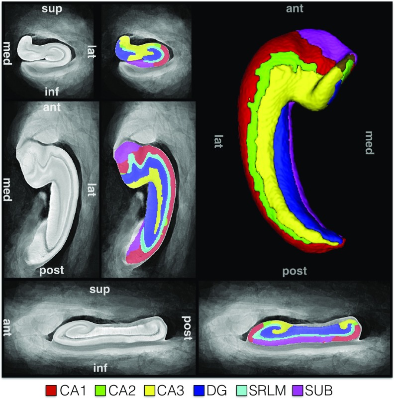
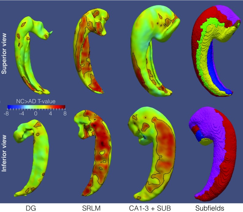
Although the hippocampus is one of the most studied structures in the human brain, limited quantitative data exist on its 3D organization, anatomical variability, and effects of disease on its subregions. Histological studies provide restricted reference information due to their 2D nature. In this paper, high-resolution (∼200 × 200 × 200 μm3) ex vivo MRI scans of 31 human hippocampal specimens are combined using a groupwise diffeomorphic registration approach into a 3D probabilistic atlas that captures average anatomy and anatomic variability of hippocampal subfields. Serial histological imaging in 9 of the 31 specimens was used to label hippocampal subfields in the atlas based on cytoarchitecture. Specimens were obtained from autopsies in patients with a clinical diagnosis of Alzheimer's disease (AD; 9 subjects, 13 hemispheres), of other dementia (nine subjects, nine hemispheres), and in subjects without dementia (seven subjects, nine hemispheres), and morphometric analysis was performed in atlas space to measure effects of age and AD on hippocampal subfields. Disproportional involvement of the cornu ammonis (CA) 1 subfield and stratum radiatum lacunosum moleculare was found in AD, with lesser involvement of the dentate gyrus and CA2/3 subfields. An association with age was found for the dentate gyrus and, to a lesser extent, for CA1. Three-dimensional patterns of variability and disease and aging effects discovered via the ex vivo hippocampus atlas provide information highly relevant to the active field of in vivo hippocampal subfield imaging.
Keywords: Alzheimer’s disease; computational anatomy; ex vivo MRI; hippocampal subfields; histology.
Conflict of interest statement
Conflict of interest statement: D.A.W. received consultation fees from Eli Lilly, Janssen, and Merck. D.A.W. receives grant support from Avid Radiopharmaceuticals/Eli Lilly, Biogen, Functional Neuromodulation, and Merck. J.B.T. has received research support from Eli Lilly. J.Q.T. may accrue revenue in the future on patents submitted by the University of Pennsylvania wherein he is co-inventor, and he received revenue from the sale of Avid to Eli Lilly as co-inventor on imaging-related patents submitted by the University of Pennsylvania.
Figures
References
-
- Duvernoy HM, et al. The Human Hippocampus. Springer; Berlin: 2005. pp. 1–232.
-
- Insausti R, Amaral DG. In: The Human Nervous System. Mai JK, Paxinos G, editors. Elsevier Academic; San Diego, Calif.: 2012.
Publication types
MeSH terms
Grants and funding
LinkOut - more resources
Full Text Sources
Other Literature Sources
Medical
Molecular Biology Databases
Miscellaneous
