

Intra-Cardiac Release of Extracellular Vesicles Shapes Inflammation Following Myocardial Infarction
- PMID: 29592957
- PMCID: PMC6023578
- DOI: 10.1161/CIRCRESAHA.117.311326
Intra-Cardiac Release of Extracellular Vesicles Shapes Inflammation Following Myocardial Infarction
Abstract
Rationale: A rapid and massive influx of inflammatory cells occurs into ischemic area after myocardial infarction (MI), resulting in local release of cytokines and growth factors. Yet, the mechanisms regulating their production are not fully explored. The release of extracellular vesicles (EVs) in the interstitial space curbs important biological functions, including inflammation, and influences the development of cardiovascular diseases. To date, there is no evidence for in situ release of cardiac EVs after MI.
Objective: The present study tested the hypothesis that local EV generation in the infarcted heart coordinates cardiac inflammation after MI.
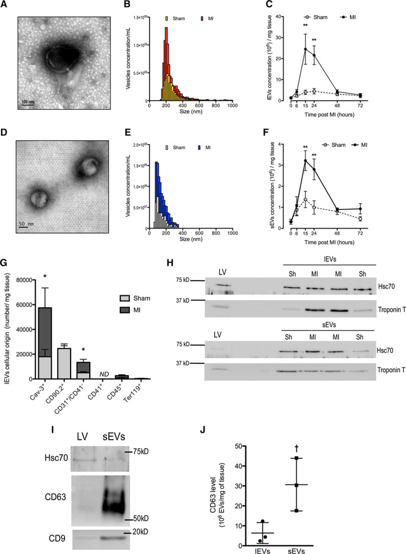
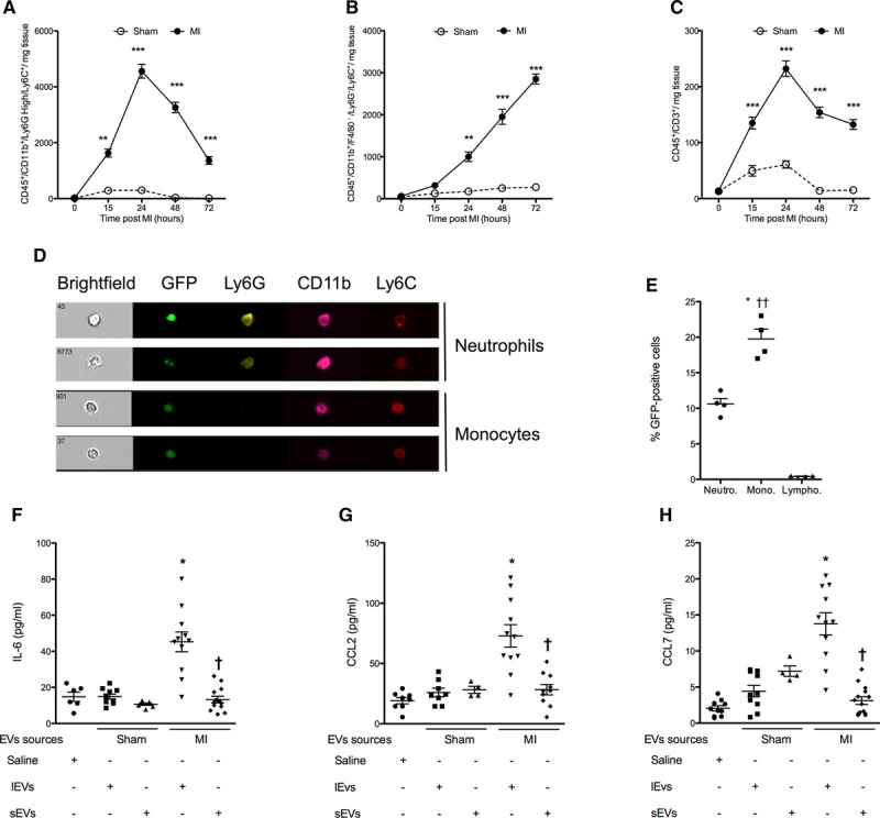
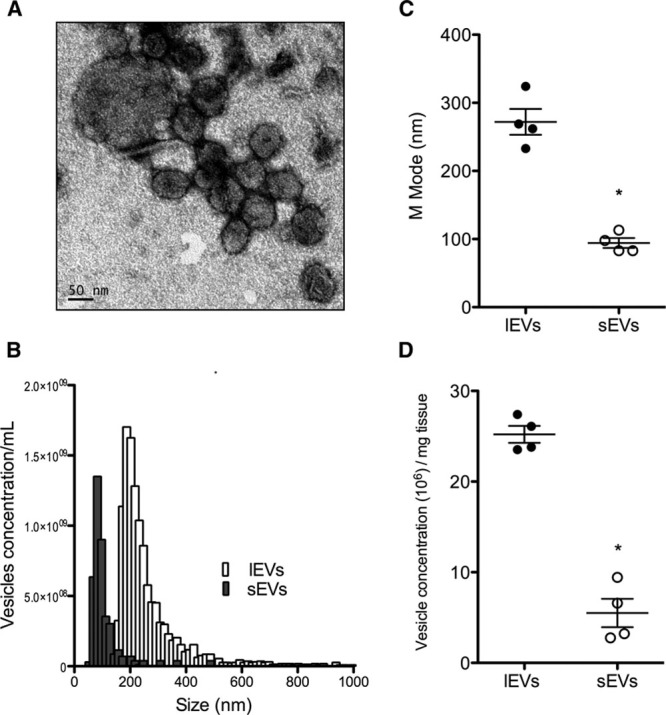
Methods and results: Coronary artery ligation in mice transiently increases EV levels in the left ventricle when compared with sham animals. EVs from infarcted hearts were characterized as large vesicles (252±18 nm) expressing cardiomyocyte and endothelial markers and small EVs (118±4 nm) harboring exosomal markers, such as CD (cluster of differentiation) 63 and CD9. Cardiac large EVs generated after MI, but not small EVs or sham EVs, increased the release of IL (interleukin)-6, CCL (chemokine ligand) 2, and CCL7 from fluorescence-activated cell-sorted Ly6C+ cardiac monocytes. EVs of similar diameter were also isolated from fragments of interventricular septum obtained from patients undergoing aortic valve replacement, thus supporting the clinical relevance of our findings in mice.
Conclusions: The present study demonstrates that acute MI transiently increases the generation of cardiac EVs characterized as both exosomes and microvesicles, originating mainly from cardiomyocytes and endothelial cells. EVs accumulating in the ischemic myocardium are rapidly taken up by infiltrating monocytes and regulate local inflammatory responses.
Keywords: exosomes; humans; inflammation; myocardial infarction; myocytes, cardiac.
© 2018 The Authors.
Figures
Comment in
-
Extracellular Vesicle Crosstalk Between the Myocardium and Immune System Upon Infarction.Circ Res. 2018 Jun 22;123(1):15-17. doi: 10.1161/CIRCRESAHA.118.313179. Circ Res. 2018. PMID: 29929970 No abstract available.
References
-
- Silvestre JS, Smadja DM, Lévy BI. Postischemic revascularization: from cellular and molecular mechanisms to clinical applications. Physiol Rev. 2013;93:1743–1802. doi: 10.1152/physrev.00006.2013. - PubMed
-
- Loyer X, Vion AC, Tedgui A, Boulanger CM. Microvesicles as cell-cell messengers in cardiovascular diseases. Circ Res. 2014;114:345–353. doi: 10.1161/CIRCRESAHA.113.300858. - PubMed
-
- Boulanger CM, Loyer X, Rautou PE, Amabile N. Extracellular vesicles in coronary artery disease. Nat Rev Cardiol. 2017;14:259–272. doi: 10.1038/nrcardio.2017.7. - PubMed
Publication types
MeSH terms
Substances
LinkOut - more resources
Full Text Sources
Other Literature Sources
Medical
