. 2018 May 29;92(12):e00017-18.
doi: 10.1128/JVI.00017-18. Print 2018 Jun 15.

Within-Host Variations of Human Papillomavirus Reveal APOBEC Signature Mutagenesis in the Viral Genome

Affiliations

Within-Host Variations of Human Papillomavirus Reveal APOBEC Signature Mutagenesis in the Viral Genome

Yusuke Hirose et al. J Virol. .

Abstract

Persistent infection with oncogenic human papillomaviruses (HPVs) causes cervical cancer, accompanied by the accumulation of somatic mutations into the host genome. There are concomitant genetic changes in the HPV genome during viral infection; however, their relevance to cervical carcinogenesis is poorly understood. Here, we explored within-host genetic diversity of HPV by performing deep-sequencing analyses of viral whole-genome sequences in clinical specimens. The whole genomes of HPV types 16, 52, and 58 were amplified by type-specific PCR from total cellular DNA of cervical exfoliated cells collected from patients with cervical intraepithelial neoplasia (CIN) and invasive cervical cancer (ICC) and were deep sequenced. After constructing a reference viral genome sequence for each specimen, nucleotide positions showing changes with >0.5% frequencies compared to the reference sequence were determined for individual samples. In total, 1,052 positions of nucleotide variations were detected in HPV genomes from 151 samples (CIN1, n = 56; CIN2/3, n = 68; ICC, n = 27), with various numbers per sample. Overall, C-to-T and C-to-A substitutions were the dominant changes observed across all histological grades. While C-to-T transitions were predominantly detected in CIN1, their prevalence was decreased in CIN2/3 and fell below that of C-to-A transversions in ICC. Analysis of the trinucleotide context encompassing substituted bases revealed that TpCpN, a preferred target sequence for cellular APOBEC cytosine deaminases, was a primary site for C-to-T substitutions in the HPV genome. These results strongly imply that the APOBEC proteins are drivers of HPV genome mutation, particularly in CIN1 lesions.IMPORTANCE HPVs exhibit surprisingly high levels of genetic diversity, including a large repertoire of minor genomic variants in each viral genotype. Here, by conducting deep-sequencing analyses, we show for the first time a comprehensive snapshot of the within-host genetic diversity of high-risk HPVs during cervical carcinogenesis. Quasispecies harboring minor nucleotide variations in viral whole-genome sequences were extensively observed across different grades of CIN and cervical cancer. Among the within-host variations, C-to-T transitions, a characteristic change mediated by cellular APOBEC cytosine deaminases, were predominantly detected throughout the whole viral genome, most strikingly in low-grade CIN lesions. The results strongly suggest that within-host variations of the HPV genome are primarily generated through the interaction with host cell DNA-editing enzymes and that such within-host variability is an evolutionary source of the genetic diversity of HPVs.

Keywords: APOBEC; genetic diversity; human papillomavirus; next-generation sequencing; quasispecies.


Figures

FIG 1
FIG 1
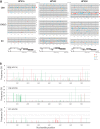
Viral whole-genome landscape of nucleotide variations observed in individual clinical specimens. (A) Positions of nucleotide variations with >0.5% frequencies of the total read count at the corresponding positions in each reference viral genome are indicated on black lines, which represent whole viral genomes of individual clinical samples. Red, orange, blue, and gray circles indicate >10%, 5 to 10%, 2 to 5%, and 0.5 to 2% frequencies, respectively. Asterisks indicate samples showing no nucleotide substitutions with >0.5% frequencies. Red stars indicate samples having a large number of C-to-T and G-to-A substitutions. The genome organizations of HPV16/52/58 are shown below: pE, the early promoter; pL, the late promoter; polyA (early) and polyA (late), the early and late polyadenylation signals, respectively. (B) Profiles of C-to-T and G-to-A substitutions throughout the viral whole genome in samples 006, 116, and 121. The frequencies of substitutions are on the y axes, whereas the x axes display nucleotide positions in the HPV genome. Red and green bars indicate C-to-T and G-to-A substitutions, respectively.
FIG 2
FIG 2
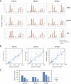
Frequency and number of nucleotide variations in the HPV genome. (A) Distribution of variation frequencies observed across the histology for HPV16/52/58. The mean frequency of nucleotide variations is indicated with a bar. (B) Boxplots of the number of nucleotide variations per sample across the histology. Statistically significant differences (P < 0.05) are indicated with asterisks.
FIG 3
FIG 3
Nonsynonymous and synonymous variations in HPV genomic regions. (A) The number of total, nonsynonymous, and synonymous nucleotide variations observed in eight viral genes (E6, E7, E1, E2, E4, E5, L2, and L1), the long control region (LCR), and a noncoding region between E5 and L2 (NC) are shown for each HPV type. (B) The correlation between the variation number and the length of nucleotide sequences in each viral genomic region. (C) Distribution of nucleotide variations between three codon positions (first, second, and third positions) in the viral open reading frames.
FIG 4
FIG 4
Nonsynonymous/nonsense variations in HPV16 E6 and E7. Amino acid variations or premature stop codons (indicated with asterisks) as within-host variations are shown in the diagrams of E6 and E7. Blue and yellow circles indicate variations observed in CIN1 and CIN2/3 samples, respectively. Stars indicate C-to-T or G-to-A substitutions. CR1, conserved region 1; CR2, conserved region 2; CR3, conserved region 3.
FIG 5
FIG 5
Mutational signature analysis of substitutions in the HPV genome. (A) Pie charts of the distribution of substitution types across the histology. All substitutions (total, n = 1,052; CIN1, n = 585; CIN2/3, n = 298; ICC, n = 169) are classified into six base substitutions, i.e., C-to-A, C-to-G, C-to-T, T-to-A, T-to-G, and T-to-G substitutions, and the relative proportion of each substitution type to all substitutions is shown in pie charts for each histological grade. (B) Distribution of 96 substitution types across the histology. All substitutions are classified into 96 base substitution types of the trinucleotide sequence context that includes the bases immediately 5′ and 3′ of each mutated base. Different colors are used to display the various types of substitutions. The relative frequencies of specific substitution types are on the y axes, whereas the x axes display different types of substitutions.
FIG 6
FIG 6
Strand specificity of C-to-T substitutions in the HPV genome. (A) Profiles of all C-to-T substitutions detected in clinical samples on each strand of HPV16/52/58 genomes. The frequencies of C-to-T substitutions are on the y axes, whereas the x axes display nucleotide positions in the HPV genome. Sense, the nontranscribed strand; antisense, the transcribed strand. N, the total number of C-to-T substitutions on the sense or antisense strand. (B) Line graphs of TpC dinucleotide density across the HPV genomes, including all lineages/sublineages for HPV16/52/58, on sense (blue line) or antisense strand (red line) of the viral genome. The gray area represents 95% confidence intervals. The genome organizations of HPV16/52/58 are shown below.

Similar articles

Cited by

References

    1. Bernard HU, Burk RD, Chen Z, van Doorslaer K, zur Hausen H, de Villiers EM. 2010. Classification of papillomaviruses (PVs) based on 189 PV types and proposal of taxonomic amendments. Virology 401:70–79. doi: 10.1016/j.virol.2010.02.002. - DOI - PMC - PubMed
    1. Bzhalava D, Eklund C, Dillner J. 2015. International standardization and classification of human papillomavirus types. Virology 476:341–344. doi: 10.1016/j.virol.2014.12.028. - DOI - PubMed
    1. Van Doorslaer K. 2013. Evolution of the papillomaviridae. Virology 445:11–20. doi: 10.1016/j.virol.2013.05.012. - DOI - PubMed
    1. Arbyn M, Tommasino M, Depuydt C, Dillner J. 2014. Are 20 human papillomavirus types causing cervical cancer? J Pathol 234:431–435. doi: 10.1002/path.4424. - DOI - PubMed
    1. zur Hausen H. 2002. Papillomaviruses and cancer: from basic studies to clinical application. Nat Rev Cancer 2:342–350. doi: 10.1038/nrc798. - DOI - PubMed

Publication types