A Vaccine Platform against Arenaviruses Based on a Recombinant Hyperattenuated Mopeia Virus Expressing Heterologous Glycoproteins
- PMID: 29593043
- PMCID: PMC5974477
- DOI: 10.1128/JVI.02230-17
A Vaccine Platform against Arenaviruses Based on a Recombinant Hyperattenuated Mopeia Virus Expressing Heterologous Glycoproteins
Abstract
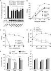
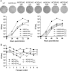
Several Old World and New World arenaviruses are responsible for severe endemic and epidemic hemorrhagic fevers, whereas other members of the Arenaviridae family are nonpathogenic. To date, no approved vaccines, antivirals, or specific treatments are available, except for Junín virus. However, protection of nonhuman primates against Lassa fever virus (LASV) is possible through the inoculation of the closely related but nonpathogenic Mopeia virus (MOPV) before challenge with LASV. We reasoned that this virus, modified by using reverse genetics, would represent the basis for the generation of a vaccine platform against LASV and other pathogenic arenaviruses. After showing evidence of exoribonuclease (ExoN) activity in NP of MOPV, we found that this activity was essential for multiplication in antigen-presenting cells. The introduction of multiple mutations in the ExoN site of MOPV NP generated a hyperattenuated strain (MOPVExoN6b) that is (i) genetically stable over passages, (ii) has increased immunogenic properties compared to those of MOPV, and (iii) still promotes a strong type I interferon (IFN) response. MOPVExoN6b was further modified to harbor the envelope glycoproteins of heterologous pathogenic arenaviruses, such as LASV or Lujo, Machupo, Guanarito, Chapare, or Sabia virus in order to broaden specific antigenicity while preserving the hyperattenuated characteristics of the parental strain. Our MOPV-based vaccine candidate for LASV, MOPEVACLASV, was used in a one-shot immunization assay in nonhuman primates and fully protected them from a lethal challenge with LASV. Thus, our hyperattenuated strain of MOPV constitutes a promising new live-attenuated vaccine platform to immunize against several, if not all, pathogenic arenaviruses.IMPORTANCE Arenaviruses are emerging pathogens transmitted to humans by rodents and responsible for endemic and epidemic hemorrhagic fevers of global concern. Nonspecific symptoms associated with the onset of infection make these viruses difficult to distinguish from other endemic pathogens. Moreover, the unavailability of rapid diagnosis in the field delays the identification of the virus and early care for treatment and favors spreading. The vaccination of exposed populations would be of great help to decrease morbidity and human-to-human transmission. Using reverse genetics, we generated a vaccine platform for pathogenic arenaviruses based on a modified and hyperattenuated strain of the nonpathogenic Mopeia virus and showed that the Lassa virus candidate fully protected nonhuman primates from a lethal challenge. These results showed that a rationally designed recombinant MOPV-based vaccine is safe, immunogenic, and efficacious in nonhuman primates.
Keywords: Lassa fever; arenavirus; innate immunity; live-vector vaccines; viral hemorrhagic fevers.
Copyright © 2018 American Society for Microbiology.
Figures


Similar articles
-
A Lassa Fever Live-Attenuated Vaccine Based on Codon Deoptimization of the Viral Glycoprotein Gene.mBio. 2020 Feb 25;11(1):e00039-20. doi: 10.1128/mBio.00039-20. mBio. 2020. PMID: 32098811 Free PMC article.
-
General Molecular Strategy for Development of Arenavirus Live-Attenuated Vaccines.J Virol. 2015 Dec;89(23):12166-77. doi: 10.1128/JVI.02075-15. Epub 2015 Sep 23. J Virol. 2015. PMID: 26401045 Free PMC article.
-
A Lassa Virus Live-Attenuated Vaccine Candidate Based on Rearrangement of the Intergenic Region.mBio. 2020 Mar 24;11(2):e00186-20. doi: 10.1128/mBio.00186-20. mBio. 2020. PMID: 32209677 Free PMC article.
-
Novel strategies for development of hemorrhagic fever arenavirus live-attenuated vaccines.Expert Rev Vaccines. 2016 Sep;15(9):1113-21. doi: 10.1080/14760584.2016.1182024. Epub 2016 May 13. Expert Rev Vaccines. 2016. PMID: 27118328 Free PMC article. Review.
-
Vaccine platforms to control Lassa fever.Expert Rev Vaccines. 2016 Sep;15(9):1135-50. doi: 10.1080/14760584.2016.1184575. Epub 2016 May 24. Expert Rev Vaccines. 2016. PMID: 27136941 Review.
Cited by
-
Depletion of CD4 and CD8 T Cells Reduces Acute Disease and Is Not Associated with Hearing Loss in ML29-Infected STAT1-/- Mice.Biomedicines. 2022 Sep 29;10(10):2433. doi: 10.3390/biomedicines10102433. Biomedicines. 2022. PMID: 36289695 Free PMC article.
-
A Lassa Fever Live-Attenuated Vaccine Based on Codon Deoptimization of the Viral Glycoprotein Gene.mBio. 2020 Feb 25;11(1):e00039-20. doi: 10.1128/mBio.00039-20. mBio. 2020. PMID: 32098811 Free PMC article.
-
Hemorrhagic Fever-Causing Arenaviruses: Lethal Pathogens and Potent Immune Suppressors.Front Immunol. 2019 Mar 13;10:372. doi: 10.3389/fimmu.2019.00372. eCollection 2019. Front Immunol. 2019. PMID: 30918506 Free PMC article. Review.
-
SAMD9L acts as an antiviral factor against HIV-1 and primate lentiviruses by restricting viral and cellular translation.PLoS Biol. 2024 Jul 3;22(7):e3002696. doi: 10.1371/journal.pbio.3002696. eCollection 2024 Jul. PLoS Biol. 2024. PMID: 38959200 Free PMC article.
-
Targeting n-myristoyltransferases promotes a pan-Mammarenavirus inhibition through the degradation of the Z matrix protein.PLoS Pathog. 2024 Dec 3;20(12):e1012715. doi: 10.1371/journal.ppat.1012715. eCollection 2024 Dec. PLoS Pathog. 2024. PMID: 39625987 Free PMC article.
References
-
- Buchmeier MJ, de la Torre J-C, Peters CJ. 2007. Arenaviridae: the viruses and their replication, p 1791–1827. In Knipe DM, Howley PM, Griffin DE, Lamb RA, Martin MA, Roizman B, Straus SE (ed), Fields virology, 5th ed Lippincott Williams & Wilkins, Philadelphia, PA.
-
- Kiley MP, Lange JV, Johnson KM. 1979. Protection of rhesus monkeys from Lassa virus by immunisation with closely related arenavirus. Lancet ii:738–745. - PubMed
Publication types
MeSH terms
Substances
Supplementary concepts
LinkOut - more resources
Full Text Sources
Other Literature Sources
Medical
Miscellaneous

