. 2018 May 29;92(12):e00197-18.
doi: 10.1128/JVI.00197-18. Print 2018 Jun 15.

Host Factor SPCS1 Regulates the Replication of Japanese Encephalitis Virus through Interactions with Transmembrane Domains of NS2B

Affiliations

Host Factor SPCS1 Regulates the Replication of Japanese Encephalitis Virus through Interactions with Transmembrane Domains of NS2B

Le Ma et al. J Virol. .

Abstract

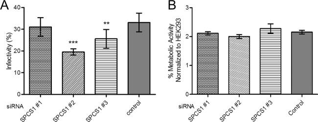
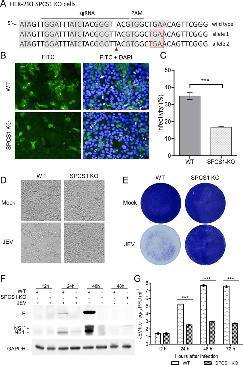
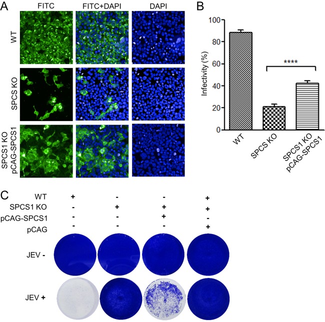
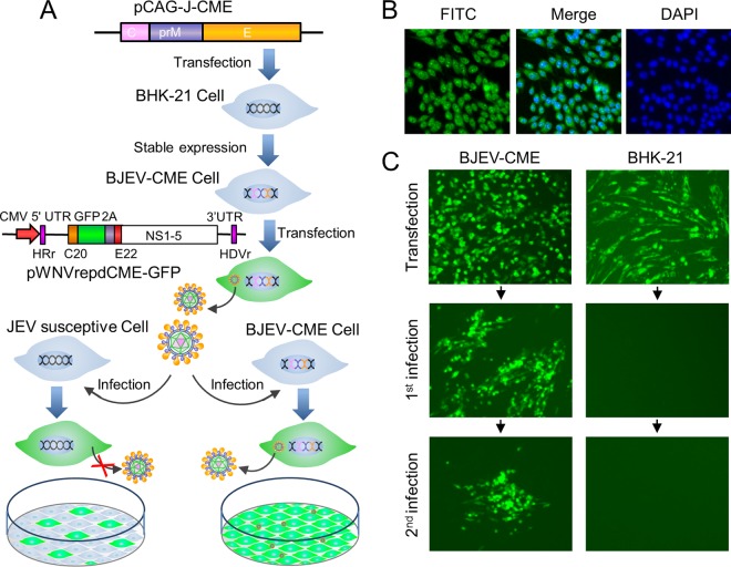
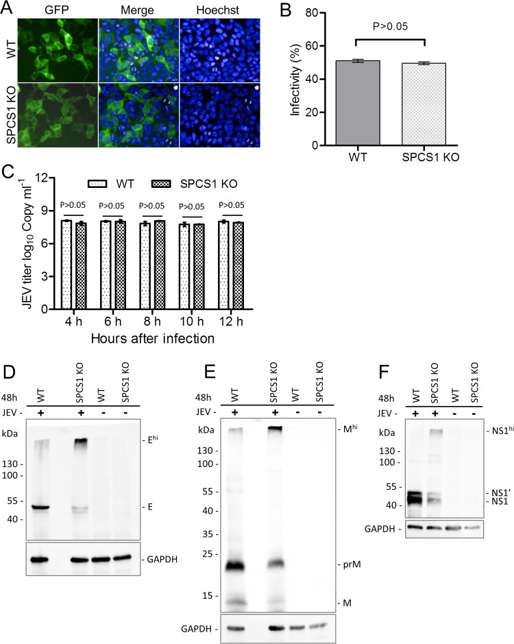
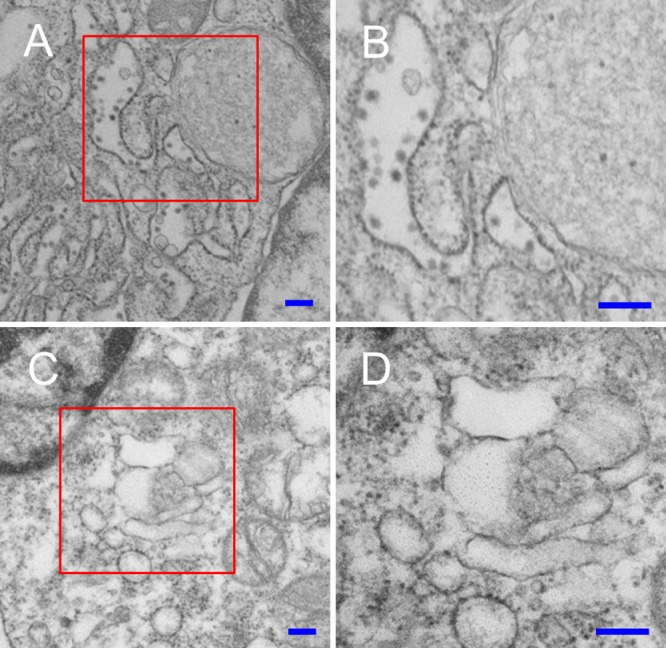
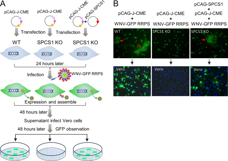
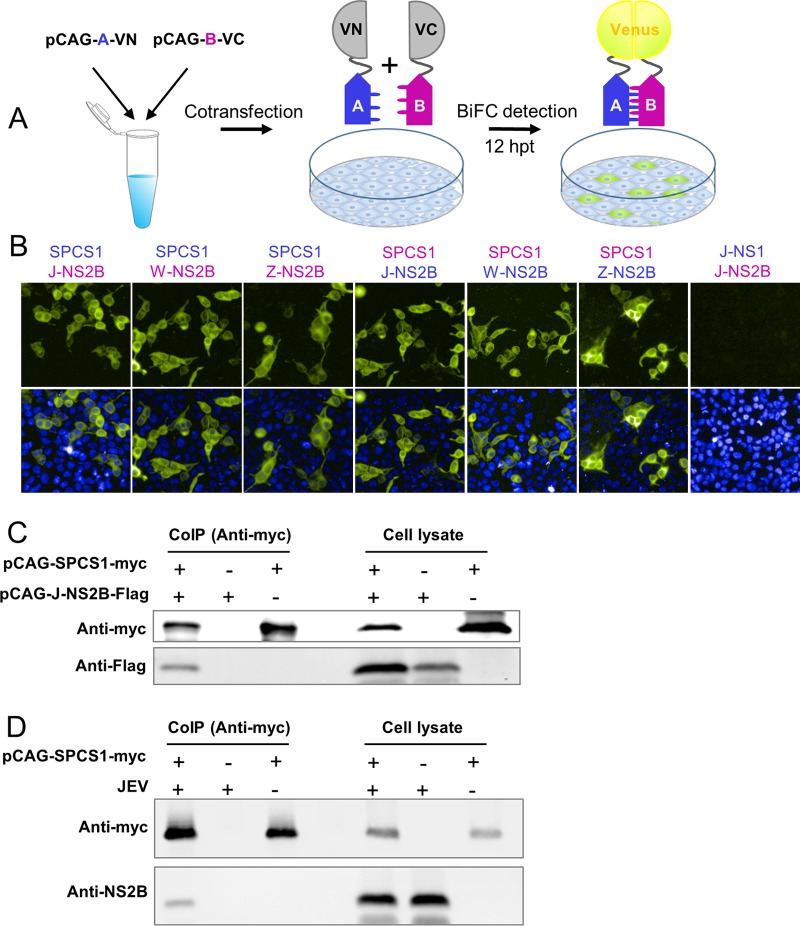
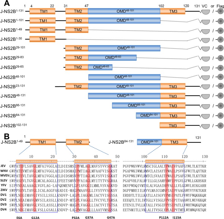
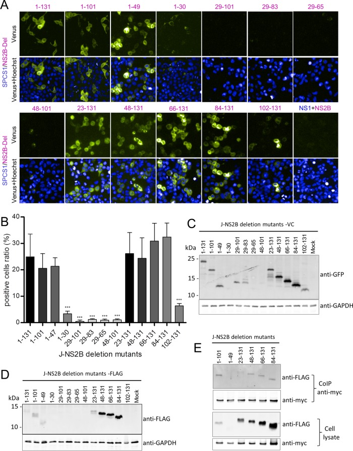
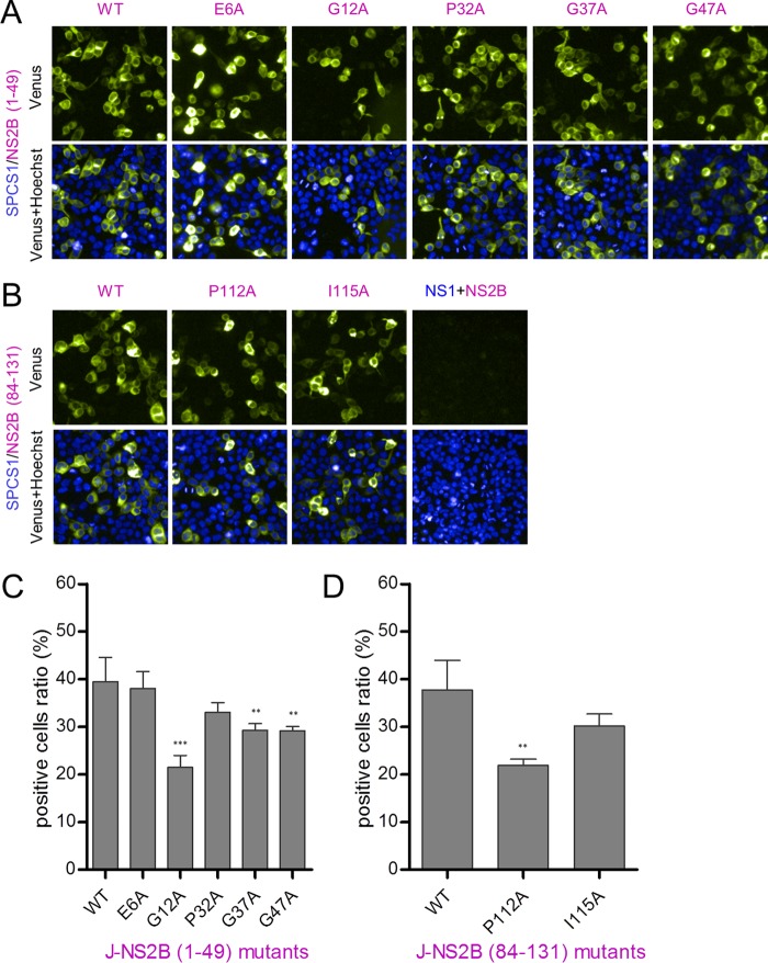
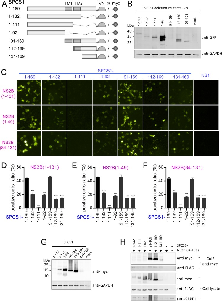
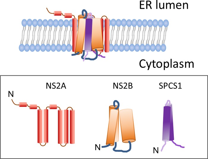
Signal peptidase complex subunit 1 (SPCS1) is a newly identified host factor that regulates flavivirus replication, but the molecular mechanism is not fully understood. Here, using Japanese encephalitis virus (JEV) as a model, we investigated the mechanism through which the host factor SPCS1 regulates the replication of flaviviruses. We first validated the regulatory function of SPCS1 in JEV propagation by knocking down and knocking out endogenous SPCS1. The loss of SPCS1 function markedly reduced intracellular virion assembly and the production of infectious JEV particles but did not affect cell entry, RNA replication, or translation of the virus. SPCS1 was found to interact with nonstructural protein 2B (NS2B), which is involved in posttranslational protein processing and virus assembly. Serial deletion mutation of the JEV NS2B protein revealed that two transmembrane domains, NS2B(1-49) and NS2B(84-131), interact with SPCS1. Further mutagenesis analysis of conserved flavivirus residues in two SPCS1 interaction domains of NS2B demonstrated that G12A, G37A, and G47A in NS2B(1-49) and P112A in NS2B(84-131) weakened the interaction with SPCS1. Deletion mutation of SPCS1 revealed that SPCS1(91-169), which contains two transmembrane domains, was involved in interactions with both NS2B(1-49) and NS2B(84-131). Taken together, these results demonstrate that SPCS1 affects viral replication by interacting with NS2B, thereby influencing the posttranslational processing of JEV proteins and the assembly of virions.IMPORTANCE Understanding virus-host interactions is important for elucidating the molecular mechanisms of virus propagation and identifying potential antiviral targets. Previous reports demonstrated that SPCS1 is involved in the flavivirus life cycle, but the mechanism remains unknown. In this study, we confirmed that SPCS1 participates in the posttranslational protein processing and viral assembly stages of the JEV life cycle but not in the cell entry, genome RNA replication, or translation stages. Furthermore, we found that SPCS1 interacts with two independent transmembrane domains of the flavivirus NS2B protein. NS2B also interacts with NS2A, which is proposed to mediate virus assembly. Therefore, we propose a protein-protein interaction model showing how SPCS1 participates in the assembly of JEV particles. These findings expand our understanding of how host factors participate in the flavivirus replication life cycle and identify potential antiviral targets for combating flavivirus infection.

Keywords: assembly; flavivirus; host factor; protein-protein interactions; viral replication.


Figures

FIG 1
FIG 1
Effect of SPCS1 knockdown on propagation of JEV. (A) HEK-293 cells were transfected with three different siRNAs targeted against SPCS1, or a control siRNA, at a final concentration of 15 nM. At 48 hpi, cells were infected with JEV at an MOI of 0.5. Two days after infection, JEV antigen-positive cells were identified by indirect immunofluorescence assays using JEV E protein-specific monoclonal antibodies. Cell nuclei were stained with 4′,6-diamidino-2-phenylindole (DAPI). Cell infectivity was examined by using an HCS system. The results are the averages of data from three independent experiments performed in triplicate. (B) Cell viability following siRNA transfection, determined by using 3-(4,5-dimethylthiazol-2-yl)-(2,5-diphenyltetrazolium bromide)-tetrazolium (MTT) cell viability assays. The data are pooled from three experiments in duplicate. Statistical significance was determined by analysis of variance with a multiple-comparison correction (**, P < 0.01; ***, P < 0.001).
FIG 2
FIG 2
Effect of the loss of SPCS1 function on propagation of JEV particles. (A) Sequencing of SPCS1 alleles in gene-edited HEK-293 cells after limiting-dilution cloning. The subgenomic RNA targeting site and protospacer adjacent motif (PAM) sequences are highlighted above the WT gene, and the sequences of edited alleles are indicated. Nucleotide triplet codons are indicated by shaded boxes. Gene editing resulting in insertions of the T nucleotide is indicated with a red arrow, and a nucleotide insertion resulting in a stop codon is indicated with a red box. (B) WT and SPCS1 KO HEK-293 cells were infected with JEV at an MOI of 0.5. At 48 hpi, cells were fixed and probed with JEV E protein-specific MAb by an immunofluorescence assay. Data from one experiment of three are shown. FITC, fluorescein isothiocyanate. (C) Cell infectivity examined with an HCS system. The data are the averages of results from three independent experiments performed in triplicate. (D and E) Cell cytopathic effects were observed at 72 hpi by microscopy (D) or crystal violet staining (E). Data from one experiment of three are shown. (F) Expression of the E and NS1 proteins in infected cells, analyzed by Western blotting with E protein- and NS1 protein-specific MAbs. Data from one experiment of two are shown. (G) Comparison of viral titers in the supernatants of WT and SPCS1 KO cells infected with JEV at an MOI of 0.01. At 12, 24, 48, and 72 hpi, the titer of infectious JEV was determined by plaque-forming assays on BHK-21 cells. The data are pooled from three experiments in duplicate. Statistical significance was determined by Student's t test (***, P < 0.001).
FIG 3
FIG 3
Effects of JEV propagation in SPCS1 KO HEK-293 cells following complementation with a plasmid expressing SPCS1. (A) WT HEK-293 cells, SPCS1 KO HEK-293 cells, and SPCS1 KO HEK-293 cells cotransfected with a plasmid expressing SPCS1 (or a control plasmid) were infected with JEV at an MOI of 0.5. At 48 hpi, cells were fixed and probed with E protein-specific monoclonal antibodies by an IFA. Data from one experiment of three are shown. (B) Infectivity analyzed by using the HCS system. The data are pooled from three experiments in duplicate. Statistical significance was determined by Student's t test (****, P < 0.0001). (C) Cell cytopathic effects in infected cells observed at 72 hpi with the crystal violet staining method. Data from one experiment of three are shown.
FIG 4
FIG 4
Generation of a cell line stably expressing the JEV C-prM-E protein and preparation of RRPs. (A) Schematic representation of the generation of a stable cell line expressing the JEV C-prM-E protein and the preparation of RRPs. The BJEV-CME cell line stably expressing the C-prM-E protein was generated by transfecting BHK-21 cells with the pCAG-opti-JEV-CME plasmid, followed by cloning and selection with G418. To produce RRPs, BJEV-CME cells were transfected with a DNA-based WNV replicon under the control of the cytomegalovirus (CMV) promoter. The replicon genome lacks the major coding sequence of the structural protein C-prM-E, and the corresponding sequence was replaced with a GFP-coding sequence following the foot-and-mouth disease virus (FMDV) 2A coding sequence. The replicon plasmid was transcribed by the cytomegalovirus promoter into the replicon RNA expressing GFP and the nonstructural replicase protein. BJEV-CME cells express the C-prM-E polyprotein, which is cleaved into the C, prM, and E proteins by replicon RNA-encoded nonstructural protease and endogenous cellular signal peptidase (SP). The replicon RNA amplifies itself again, and the three structural proteins package the replicon RNA into RRPs, which are secreted into the culture medium. When RRPs infect other JEV-susceptible cells, such as BHK-21 cells, the replicon RNA expresses GFP and nonstructural proteins, which amplifies more RNA. However, no structural proteins or additional RRPs are produced in RRP-infected cells, thereby preventing further replication. When BJEV-CME cells are infected with RRPs, the structural proteins expressed by the cells package the replicon RNA into progeny RRPs, and the infection spreads in rounds similar to those for the wild-type virus. UTR, untranslated region. (B) Cloned and selected stable cell lines were identified by an IFA with JEV E protein-specific MAb 5E7. (C) Production of RRPs from the WNV replicon plasmid by transfection of BJEV-CME cell lines. Green florescence was visualized when replicon plasmid pWNVrepdCME-GFP was transfected into BJEV-CME cells (first infection) and BHK-21 cells (second infection). Transfected-cell supernatants were passaged onto fresh cell cultures, as indicated by the arrows, and green florescence was observed only in BJEV-CME cells. At 3 days postinoculation, supernatants of cells infected first were used to inoculate cells, as indicated by the arrows, for a second infection, and green florescence was analyzed at 72 h postinfection. HRr, hammerhead ribozyme; HDVr, hepatitis delta virus ribozyme.
FIG 5
FIG 5
Effects of the loss of SPCS1 function on virus entry into cells, genome RNA replication, and protein processing during JEV infection. (A) WT and SPCS1 KO HEK-293 cells were infected with JEV single-round infectious RRPs at an MOI of 0.5. At 48 hpi, cell nuclei were stained with Hoechst reagent. Cells positive for GFP fluorescence indicated infection with RRPs. Data from one experiment of three are shown. (B) Infectivity analyzed by using the HCS system. The data are pooled from three experiments in duplicate. Statistical significance was determined by Student's t test (*, P < 0.05). (C) Real-time qRT-PCR analysis of JEV during the early stages of infection. The data are pooled from three experiments in duplicate. Statistical significance was determined by Student's t test (*, P < 0.05). (D to F) WT and SPCS1 KO HEK-293 cells were infected with JEV at an MOI of 1.0. At 4, 6, 8, 10, and 12 hpi, infected cells were harvested, and JEV genome RNAs were analyzed by qRT-PCR. To examine the expression of the JEV E, prM, and NS1 proteins in infected cells, WT and SPCS1 KO HEK-293 cells were infected with JEV at an MOI of 20. At 48 hpi, cell lysates were blotted with anti-JEV E protein (D), anti-JEV prM protein (E), or anti-JEV NS1 protein (F) monoclonal antibodies. Higher-molecular-mass bands (Ehi, Mhi, and NS1hi) reacting with the respective monoclonal antibodies are indicated. Data from one experiment of two are shown.
FIG 6
FIG 6
Electron micrographs of WT and SPCS1 KO HEK-293 cells infected with JEV. (A) Low-magnification image of JEV-infected WT HEK-293 cells. (B) Enlargement of the area boxed in red in panel A. Virus particles in the ER are indicated by red arrowheads. (C) Low-magnification image of JEV-infected SPCS1 KO HEK-293 cells. (D) Enlargement of the area boxed in red in panel C. Bars = 200 nm.
FIG 7
FIG 7
Effects of the loss of SPCS1 function on virus assembly. (A) Schematic diagram of the experimental workflow. SPCS1 KO HEK-293 cells were transfected with plasmid pCAG-J-CME expressing JEV structural proteins with or without plasmid pCAG-SPCS1 expressing SPCS1. WT HEK-293 cells were transfected with pCAG-J-CME as positive controls. Transfected cells were infected with WNV-GFP RRPs (21) at 24 hpi. At 48 hpi, supernatants were harvested and used to infect Vero cells. GFP expression in cells was observed by fluorescence microscopy at 48 hpi. (B) Visualization of GFP expression following transfection with plasmids and infection with WNV-GFP RRPs (top row) and in supernatants of infected Vero cells (bottom row). Progeny viruses represented by the presence of GFP foci are indicated with red arrows.
FIG 8
FIG 8
Interaction of SPCS1 with the JEV NS2B protein in mammalian cells. (A) Schematic diagram of BiFC analysis. Blue A's and purple B's represent a pair of proteins analyzed by BiFC. VN and VC represent the N-terminal fragment (residues 1 to 173) and the C-terminal fragment (residues 174 to 239) of the Venus protein, respectively. hpt, hours posttransfection. (B) Detection of the interaction of SPCS1-VN with VC fused to the NS2B proteins of JEV, WNV, and ZIKV and the corresponding reverse interactions of SPCS1-VC with VN fused to the NS2B proteins of JEV, WNV, and ZIKV. Data from one experiment of three are shown. (C) HEK-293T cells were cotransfected with separate plasmids expressing SPCS1-myc and FLAG-tagged NS2B. Cell lysates of transfected cells were immunoprecipitated with anti-myc antibody. The resulting precipitates and whole-cell lysates used for immunoprecipitation were examined by immunoblotting using anti-myc and anti-FLAG antibodies. Data from one experiment of two are shown. CoIP, coimmunoprecipitation. (D) HEK-293T cells were transfected with a plasmid expressing SPCS1-myc. Cells were infected with JEV at an MOI of 0.5 at 24 h posttransfection. At 48 hpi, cell lysates of infected cells were immunoprecipitated with anti-myc antibody. The resulting precipitates and whole-cell lysates used for immunoprecipitation were examined by immunoblotting using anti-myc and anti-NS2B antibodies. Data from one experiment of two are shown.
FIG 9
FIG 9
Schematic diagram of JEV NS2B deletion mutant constructs and sequence alignment of the transmembrane domains of flavivirus NS2B proteins. (A) JEV NS2B deletion mutant constructs tagged with the C-terminal fragment of the Venus protein or FLAG. Transmembrane segments are indicated in orange boxes. The main outer membrane domains (OMD) are indicated in blue boxes. The locations of NS2B deletion mutants are indicated on the left, with amino acid positions in superscript type. (B) Alignment of flavivirus NS2B transmembrane domain sequences. Conserved residues between groups are shaded in red. Five conserved residues in the TM1 and TM2 regions, and two conserved residues in the TM3 region, were selected for site-directed mutagenesis. For point mutations, all selected amino acid residues were replaced with alanine. MVEV, Murray Valley encephalitis virus; SLEV, Saint Louis encephalitis virus; DV1 to DV4, dengue virus 1 to 4.
FIG 10
FIG 10
Interaction of SPCS1 with JEV NS2B deletion mutants. (A) BiFC analysis of the interaction between SPCS1-VN and VC fused to JEV NS2B or its deletion mutants. Images were taken at 12 hpi by using the HCS system. Data from one experiment of three are shown. (B) The ratio of positive cells was determined by using the HCS system. The data are pooled from three experiments in duplicate. Statistical significance was determined by analysis of variance with a multiple-comparison correction (***, P < 0.001). (C) Expression of BiFC constructs. HEK-293T cells were transfected with NS2B deletion mutants tagged with VC. Cell lysates were examined by immunoblotting using anti-GFP or anti-GAPDH antibodies. Data from one experiment of two are shown. (D) Expression of FLAG-tagged JEV NS2B deletion mutants. HEK-293T cells were transfected with the indicated plasmids, and cell lysates were examined by immunoblotting using anti-FLAG or anti-GAPDH antibodies. Data from one experiment of two are shown. (E) Cells were cotransfected with the indicated NS2B deletion mutant plasmids, and lysates of transfected cells were immunoprecipitated with anti-myc antibody. The resulting precipitates and whole-cell lysates used for immunoprecipitation were examined by immunoblotting using anti-FLAG or anti-myc antibodies. Data from one experiment of two are shown.
FIG 11
FIG 11
Interaction of SPCS1 with point mutants of JEV NS2B(1–49) and NS2B(84–131). (A and B) Visualization of the interaction between SPCS1-VN and VC fused with point mutants of JEV NS2B(1–49) (A) and NS2B(84–131) (B) using the BiFC system. Images were taken at 12 hpi by using the HCS system. Data from one of two independent experiments performed in triplicate are shown. (C and D) The ratios of positive cells were determined. Shown are data from analyses of the statistical significance of differences between point mutants and the corresponding WT NS2B(1–49) (C) and NS2B(84–131) (D). Data are the averages of results from two independent experiments performed in triplicate. Statistical significance was determined by analysis of variance with a multiple-comparison correction (**, P < 0.01; ***, P < 0.001).
FIG 12
FIG 12
Interaction of SPCS1 deletion mutants with JEV NS2B, NS2B(1–49), and NS2B(84–131). (A) Schematic diagram of SPCS1 deletion mutant constructs tagged with the N-terminal fragment of the Venus protein or myc. Transmembrane segments are indicated in gray boxes. The locations of SPCS1 deletion mutants are indicated on the left, along with amino acid positions. (B) Expression of VN-tagged SPCS1 deletion mutants. The membrane was probed with mouse monoclonal antibodies against GFP. Data from one experiment of two are shown. (C) Plasmids were cotransfected into HEK-293T cells, images were captured at 12 hpi by using the HCS system, and the ratio of positive cells was determined. Data from one experiment of three are shown. (D) Positive cell ratios for the interaction between SPCS1 deletion mutants and JEV NS2B. (E) Positive cell ratios for the interaction between SPCS1 deletion mutants and JEV NS2B(1–49). (F) Positive cell ratios for the interaction between SPCS1 deletion mutants and JEV NS2B(84–131). The data are pooled from three experiments in duplicate. Statistical significance was determined by analysis of variance with a multiple-comparison correction (***, P < 0.001). (G) Expression of myc-tagged SPCS1 deletion mutants. HEK-293T cells were transfected with the indicated plasmids, and cell lysates were examined by immunoblotting using anti-myc or anti-GAPDH antibodies. Data from one experiment of two are shown. (H) Cells were cotransfected with JEV NS2B(84–131) and the indicated SPCS1 deletion mutants, and lysates of transfected cells were immunoprecipitated with anti-myc antibody. The resulting precipitates and whole-cell lysates used for immunoprecipitation were examined by immunoblotting using anti-FLAG or anti-myc antibodies. Data from one experiment of two are shown.
FIG 13
FIG 13
Proposed model for the interaction of NS2B with SPCS1 and NS2A.

Similar articles

Cited by

References

    1. Chambers TJ, Hahn CS, Galler R, Rice CM. 1990. Flavivirus genome organization, expression, and replication. Annu Rev Microbiol 44:649–688. doi:10.1146/annurev.mi.44.100190.003245. - DOI - PubMed
    1. Mukhopadhyay S, Kuhn RJ, Rossmann MG. 2005. A structural perspective of the flavivirus life cycle. Nat Rev Microbiol 3:13–22. doi:10.1038/nrmicro1067. - DOI - PubMed
    1. Leung JY, Pijlman GP, Kondratieva N, Hyde J, Mackenzie JM, Khromykh AA. 2008. Role of nonstructural protein NS2A in flavivirus assembly. J Virol 82:4731–4741. doi:10.1128/JVI.00002-08. - DOI - PMC - PubMed
    1. Miller S, Kastner S, Krijnse-Locker J, Buhler S, Bartenschlager R. 2007. The non-structural protein 4A of dengue virus is an integral membrane protein inducing membrane alterations in a 2K-regulated manner. J Biol Chem 282:8873–8882. doi:10.1074/jbc.M609919200. - DOI - PubMed
    1. Umareddy I, Chao A, Sampath A, Gu F, Vasudevan SG. 2006. Dengue virus NS4B interacts with NS3 and dissociates it from single-stranded RNA. J Gen Virol 87:2605–2614. doi:10.1099/vir.0.81844-0. - DOI - PubMed

Publication types

MeSH terms

Substances