. 2018 May 29;92(12):e02011-17.
doi: 10.1128/JVI.02011-17. Print 2018 Jun 15.

eCD4-Ig Variants That More Potently Neutralize HIV-1

Affiliations

eCD4-Ig Variants That More Potently Neutralize HIV-1

Ina Fetzer et al. J Virol. .

Abstract

The human immunodeficiency virus type 1 (HIV-1) entry inhibitor eCD4-Ig is a fusion of CD4-Ig and a coreceptor-mimetic peptide. eCD4-Ig is markedly more potent than CD4-Ig, with neutralization efficiencies approaching those of HIV-1 broadly neutralizing antibodies (bNAbs). However, unlike bNAbs, eCD4-Ig neutralized all HIV-1, HIV-2, and simian immunodeficiency virus (SIV) isolates that it has been tested against, suggesting that it may be useful in clinical settings, where antibody escape is a concern. Here, we characterize three new eCD4-Ig variants, each with a different architecture and each utilizing D1.22, a stabilized form of CD4 domain 1. These variants were 10- to 20-fold more potent than our original eCD4-Ig variant, with a construct bearing four D1.22 domains (eD1.22-HL-Ig) exhibiting the greatest potency. However, this variant mediated less efficient antibody-dependent cell-mediated cytotoxicity (ADCC) activity than eCD4-Ig itself or several other eCD4-Ig variants, including the smallest variant (eD1.22-Ig). A variant with the same architecture as the original eCD4-Ig (eD1.22-D2-Ig) showed modestly higher thermal stability and best prevented the promotion of infection of CCR5-positive, CD4-negative cells. All three variants, and eCD4-Ig itself, mediated more efficient shedding of the HIV-1 envelope glycoprotein gp120 than did CD4-Ig. Finally, we show that only three D1.22 mutations contributed to the potency of eD1.22-D2-Ig and that introduction of these changes into eCD4-Ig resulted in a variant 9-fold more potent than eCD4-Ig and 2-fold more potent than eD1.22-D2-Ig. These studies will assist in developing eCD4-Ig variants with properties optimized for prophylaxis, therapy, and cure applications.IMPORTANCE HIV-1 bNAbs have properties different from those of antiretroviral compounds. Specifically, antibodies can enlist immune effector cells to eliminate infected cells, whereas antiretroviral compounds simply interfere with various steps in the viral life cycle. Unfortunately, HIV-1 is adept at evading antibody recognition, limiting the utility of antibodies as a treatment for HIV-1 infection or as part of an effort to eradicate latently infected cells. eCD4-Ig is an antibody-like entry inhibitor that closely mimics HIV-1's obligate receptors. eCD4-Ig appears to be qualitatively different from antibodies, since it neutralizes all HIV-1, HIV-2, and SIV isolates. Here, we characterize three new structurally distinct eCD4-Ig variants and show that each excels in a key property useful to prevent, treat, or cure an HIV-1 infection. For example, one variant neutralized HIV-1 most efficiently, while others best enlisted natural killer cells to eliminate infected cells. These observations will help generate eCD4-Ig variants optimized for different clinical applications.

Keywords: CD4; CD4-Ig; coreceptor; eCD4-Ig; human immunodeficiency virus.


Figures

FIG 1
FIG 1
Architecture of eCD4-Ig variants. eCD4-Ig and variants including the D1.22 domain described by Chen et al. (23) are presented. Light blue, native CD4 immunoglobulin domains 1 and 2; dark blue, the D1.22 variant of CD4 domain 1 with six amino acid differences from native domain 1; gray, the Fc domain of human IgG1; red, the tyrosine-sulfated coreceptor-mimetic peptide mim6 (sequence: GGGGGDYYDYDGGYYYDGD). (Left to right) Original eCD4-Ig, a fusion of CD4 domains 1 and 2, the Fc region, and mim6 (19); eD1.22-Ig, with CD4 domains 1 and 2 of eCD4-Ig replaced by D1.22, a stabilized CD4 domain 1; eD1.22-D2-Ig, in which the D1.22 domain replaces CD4 domain 1 but CD4 domain 2 of eCD4-Ig is retained; eD1.22-HL-Ig, an antibody-like construct in which D1.22 domains replace the variable regions of an IgG1 heavy chain and a kappa light chain. As with eCD4-Ig, mim6 is fused to the C terminus of the heavy chain.
FIG 2
FIG 2
Stabilizing mutations in CD4 domain 1 improve the neutralization efficiency of eCD4-Ig variants. (A) The neutralization efficiencies of the indicated eCD4-Ig variants or the bNAb 10-1074 were determined using a TZM-bl neutralization assay. The indicated variants were preincubated for 1 h with HIV-1 pseudotyped with the Env proteins of 19 diverse HIV-1 isolates. TZM-bl cells were added and incubated for 48 to 72 h. Luciferase expression was measured and normalized to expression in the absence of any inhibitor. IC50s were plotted. Geometric means for neutralized isolates are indicated by horizontal lines. The numbers of isolates resistant to 50 μg/ml are indicated at the top. All comparisons with eCD4-Ig and eD1.22-HL-Ig were significant (P < 0.01; paired Student's t test). (B) Representative neutralization studies used to generate panel A. VSV-G indicates HIV-1 pseudotyped with the VSV-G entry protein. The error bars indicate standard errors of the mean (SEM) of triplicates.
FIG 3
FIG 3
IC50s of eCD4-Ig variants against diverse HIV-1 isolates. The IC50s of CD4-Ig, eCD4-Ig, three eCD4-Ig variants, and 10-1074 are listed for each of the isolates shown in Fig. 2A, with the clades of the isolates indicated. The colors indicate the IC50 ranges.
FIG 4
FIG 4
Expression efficiencies and aggregation temperatures of eCD4-Ig variants. (A) Average expression yields of eCD4-Ig variants from transiently transfected Expi293 cells. The eCD4-Ig variants were purified with protein A columns, and protein concentrations were determined by absorbance measurement at a wavelength of 280 nm. (B) The thermal stability of eCD4-Ig variants was determined by DLS. The hydrodynamic radius of each protein was measured as temperatures increased. Shown are the temperatures at which aggregation, defined as a marked increase of radius, was observed. The error bars indicate the SEM for the results of three independent experiments.
FIG 5
FIG 5
ADCC activities of eCD4-Ig variants. The ADCC activities of different eCD4-Ig variants or the bNAbs PGT121 and 3BNC117 were determined using CEM.NKR-CCR5-LTR-Luc cells that were infected with HIV-1 isolate YU2, NL4-3, or 89.6, as indicated. Three or 4 days postinfection, cells in a 10:1 effector-to-target ratio were added in the presence of the indicated concentrations of the eCD4-Ig variants. ADCC responses, defined as a loss of luciferase signal in relative light units (RLU), were measured after an 8-h incubation. The error bars indicate SEM of triplicates. The data are representative of at least two independent experiments with similar results.
FIG 6
FIG 6
eCD4-Ig variants vary in their abilities to promote infection of CD4-negative cells. The ability of eCD4-Ig to promote infection of CD4-negative, CCR5-positive cells was determined using viruses pseudotyped with the Env proteins of the X1632, CE1176, CH119, and YU2 isolates. Pseudoviruses pseudotyped with these Envs and modified to express firefly luciferase were preincubated with eCD4-Ig for 1 h and then mixed with Cf2Th-CCR5 cells for 48 to 72 h. Luciferase expression was plotted as a percentage of the maximum value observed for each pseudovirus. The error bars represent SEM of triplicates. The data are representative of at least two independent experiments with similar results. Note that neither eD1.22-D2-Ig nor eCD4-Ig detectably enhanced infection. Area-under-the-curve comparisons of CD4-Ig and eD1.22-Ig with eCD4-Ig and eD1.22-D2-Ig were significant for the isolates X1632, CE1176, and CH119 (P < 0.03; unpaired t test). Comparisons between eD1.22-HL-Ig, eCD4-Ig, and eD1.22-D2-Ig were significant only for the X1632 isolate.
FIG 7
FIG 7
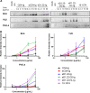
eCD4-Ig variants promote more efficient dissociation of gp120 from gp41 than does CD4-Ig. (A) HEK293T cells were transiently transfected with plasmids encoding the Env proteins of the 89.6, YU2, and PVO.4 HIV-1 isolates. Two days later, the cells were incubated with the indicated concentrations of CD4-Ig, eCD4-Ig, eD1.22-Ig, eD1.22-D2-Ig, eD1.22-HL-Ig, or the bNAb 10-1074. After 1 h, the supernatants were removed, the proteins were separated by SDS-PAGE, and the gels were analyzed by Western blotting with sheep anti-gp120 polyclonal serum. The data are representative of three independent experiments with similar results. (B) gp120 bands were quantified from phosphoimages using ImageJ software and normalized to densities observed without protein incubation. The error bars indicate SEM of the results of three independent experiments.
FIG 8
FIG 8
An eCD4-Ig variant bearing 3 of 6 D1.22 mutations is more potent than eCD4-Ig or eD1.22-D2-Ig. (A) Expression yields were measured as for Fig. 4A, except that eCD4-Ig3mut, an eCD4-Ig variant bearing 3 of 6 D1.22 mutations, was compared to eCD4-Ig and eD1.22-D2-Ig. (B) DLS experiment similar to that shown in Fig. 4B, except that, again, eCD4-Ig3mut was characterized. (C) Summary of neutralization studies as shown in Fig. 2, except that eCD4-Ig, eD1.22-D2-Ig, and eCD4-Ig3mut were compared. Note that eCD4-Ig3mut is 2-fold and 9-fold more potent than eD1.22-D2-Ig and eCD4-Ig, respectively. Both comparisons with eCD4-Ig3mut were statistically significant (P = 0.016 with eCD4-Ig and P = 0.012 with eD1.22-D2-Ig; paired Student's t test).

References

    1. Checkley MA, Luttge BG, Freed EO. 2011. HIV-1 envelope glycoprotein biosynthesis, trafficking, and incorporation. J Mol Biol 410:582–608. doi:10.1016/j.jmb.2011.04.042. - DOI - PMC - PubMed
    1. Wilen CB, Tilton JC, Doms RW. 2012. HIV: cell binding and entry. Cold Spring Harb Perspect Med 2:a006866. doi:10.1101/cshperspect.a006866. - DOI - PMC - PubMed
    1. Liu J, Bartesaghi A, Borgnia MJ, Sapiro G, Subramaniam S. 2008. Molecular architecture of native HIV-1 gp120 trimers. Nature 455:109–113. doi:10.1038/nature07159. - DOI - PMC - PubMed
    1. Choe H, Farzan M, Sun Y, Sullivan N, Rollins B, Ponath PD, Wu L, Mackay CR, LaRosa G, Newman W, Gerard N, Gerard C, Sodroski J. 1996. The beta-chemokine receptors CCR3 and CCR5 facilitate infection by primary HIV-1 isolates. Cell 85:1135–1148. doi:10.1016/S0092-8674(00)81313-6. - DOI - PubMed
    1. Feng Y, Broder CC, Kennedy PE, Berger EA. 1996. HIV-1 entry cofactor: functional cDNA cloning of a seven-transmembrane, G protein-coupled receptor. Science 272:872–877. doi:10.1126/science.272.5263.872. - DOI - PubMed

Publication types

MeSH terms