Review
. 2018 Mar 1;32(5-6):327-340.
doi: 10.1101/gad.312561.118.

Necroptosis in development and diseases

Affiliations
Review

Necroptosis in development and diseases

Bing Shan et al. Genes Dev. .

Abstract

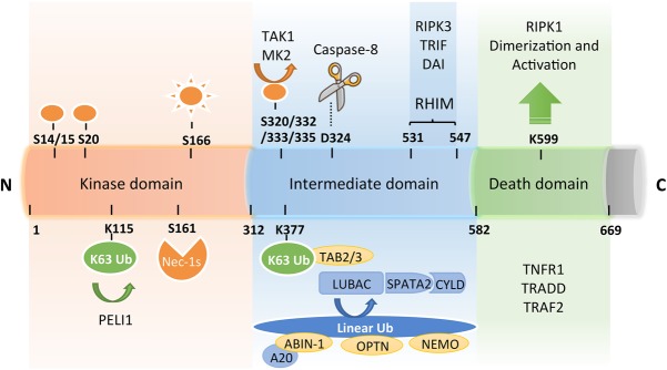
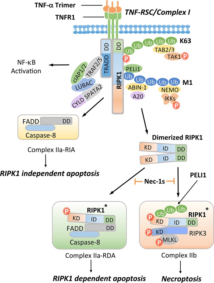
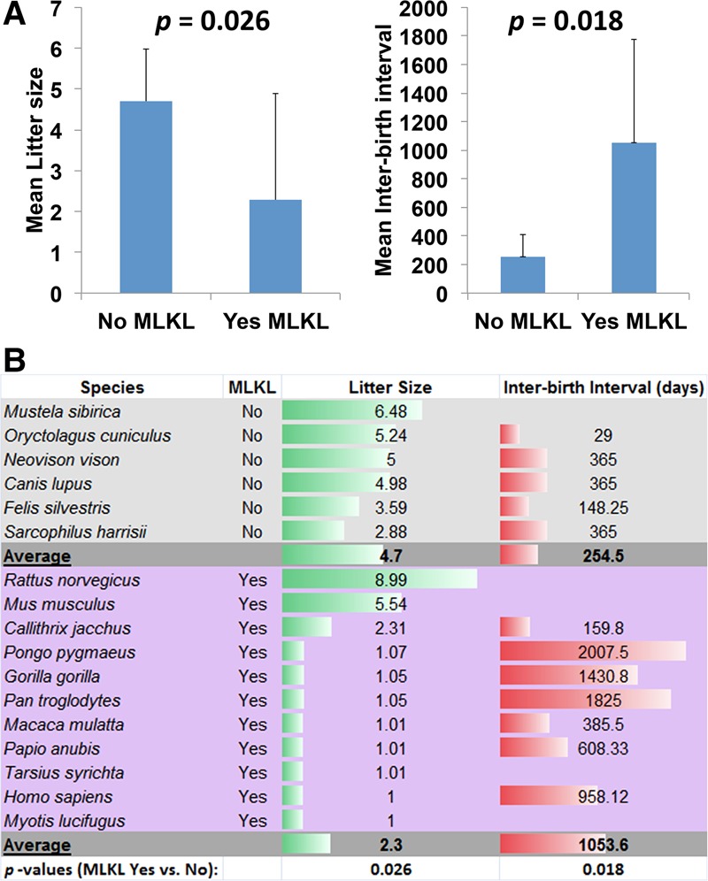
Necroptosis, a form of regulated necrotic cell death mediated by RIPK1 (receptor-interacting protein kinase 1) kinase activity, RIPK3, and MLKL (mixed-lineage kinase domain-like pseudokinase), can be activated under apoptosis-deficient conditions. Modulating the activation of RIPK1 by ubiquitination and phosphorylation is critical to control both necroptosis and apoptosis. Mutant mice with kinase-dead RIPK1 or RIPK3 and MLKL deficiency show no detrimental phenotype in regard to development and adult homeostasis. However, necroptosis and apoptosis can be activated in response to various mutations that result in the abortion of the defective embryos and human inflammatory and neurodegenerative pathologies. RIPK1 inhibition represents a key therapeutic strategy for treatment of diseases where blocking both necroptosis and apoptosis can be beneficial.

Keywords: MLKL; RIPK1; RIPK3; apoptosis; necroptosis.


Figures

Figure 1.
Figure 1.
A schematic diagram of RIPK1 domains, interacting proteins, post-translational modifications, and the catalytic enzymes that read or write these modifications. RIPK1 contains an N-terminal kinase domain, an intermediate domain with a RIP homotypic interaction motif (RHIM), and a C-terminal DD. The phosphorylation and ubiquitination sites as well as types of ubiquitin linkages on RIPK1 are indicated together with their respective catalytic enzymes. K63 ubiquitination chains on Lys377 mediate the recruitment of TAB2/3 and the activation of transforming growth factor-β-activated kinase 1 (TAK1), which in turn phosphorylates IKKs for activating the NF-κB pathway. M1 ubiquitination of RIPK1 is regulated by the linear ubiquitination assembly complex (LUBAC) and the deubiqutinating complex CYLD/SPATA2. The ubiquitin-binding proteins ABIN-1, Optineurin (OPTN), and NEMO, each carrying a UBAN motif that can bind with the linear ubiquitination chains, play important roles in regulating the activation of RIPK1. The RHIM motif of RIPK1 is required for binding with RIPK3 to mediate necroptosis and may also interact with TRIF, DAI, and Toll-like receptors to promote inflammation. Autophosphorylation of Ser166 is a biomarker for RIPK1 activation. The small molecule Nec-1s is caged in a hydrophobic pocket between the N and C lobes of the kinase domain and forms an H bond between its nitrogen atom and the hydroxyl oxygen of Ser161 on the activation loop to inhibit the activation of RIPK1. The cleavage of RIPK1 after Asp324 by caspase-8 or the phosphorylation on Ser320/331/333/335 in human RIPK1 and Ser321/332/334/336 in murine RIPK1 by TAK1 or MK2 leads to the suppression of RIPK1 activation. E3 ligase Pellino 1 (PELI1) mediates K63 ubiquitination on Lys115 of activated RIPK1 to promote complex IIb formation and necroptosis. The DD is not only crucial for the initiation of TNFR1 signaling but also indispensable for RIPK1 activation by mediating RIPK1 dimerization during the transition from complex I to complex II.
Figure 2.
Figure 2.
Activation of TNFR1 may promote multiple alternative signaling pathways, including the activation of NF-κB, RIPK1-independent apoptosis (RIA), RDA, and necroptosis. Activation of TNFR1 by trimerized TNFα induces the formation of a membrane-associated transient complex (named complex I or TNF-RSC), which includes RIPK1, TRADD, TRAF2/5, cIAP1/2, LUBAC, ABIN-1, A20, NEMO, PELI1, CYLD, and SPATA2. In complex I, RIPK1 is rapidly polyubiquitinated by K63-linked and M1-linked ubiquitin chains mediated by cIAP1/2 and LUBAC, respectively. K63 ubiquitination chains on RIPK1 mediate the recruitment of TAB2/3 to facilitate the activation of TAK1, leading to phosphorylation of the IKK complex and RIPK1 on S320/S331/S333/S335. M1 ubiquitination chains on RIPK1 mediate the recruitment of PELI1, ABIN-1, and NEMO. ABIN-1 in turn recruits phosphorylated A20 to promote K63 deubiquitination of RIPK1. CYLD/SPATA2 promote the M1 deubiquitination of RIPK1. The activated IKK/NEMO complex promotes the activation of NF-κB. When the output of the NF-κB pathway is inhibited by cycloheximide, which blocks the expression of cFLIPL, FADD interacts with caspase-8 to form complex IIa-RIA to promote RIA. Alternatively, dimerization of RIPK1 can mediate the activation of RIPK1 during the transition from complex I to complex II. Activated RIPK1* can interact with FADD and caspase-8 to form complex IIa-RDA to promote RDA. When caspase-8 is inhibited, activated RIPK1* is ubiquitinated by PELI1 on K115 and binds with RIPK3 to form complex IIb, leading to MLKL activation and necroptosis. Nec-1s inhibits the activation of RIPK1 kinase and in turn prevents the formation of complex IIa to block RDA and complex IIb to block necroptosis.
Figure 3.
Figure 3.
MLKL expression and necroptosis in fast- and slow-breeding mammalian species. (A) Comparison of litter sizes and interbirth intervals in mammals that express or do not express MLKL. Mean values are shown, and error bars represent standard deviation. Student's t-test was used to calculate the P-values. (B) List of species used in A and their mean litter sizes and mean interbirth intervals.

References

    1. Aggarwal BB. 2003. Signalling pathways of the TNF superfamily: a double-edged sword. Nat Rev Immunol 3: 745–756. - PubMed
    1. Aigelsreiter A, Haybaeck J, Schauer S, Kiesslich T, Bettermann K, Griessbacher A, Stojakovic T, Bauernhofer T, Samonigg H, Kornprat P, et al. 2012. NEMO expression in human hepatocellular carcinoma and its association with clinical outcome. Hum Pathol 43: 1012–1019. - PubMed
    1. Alcamo E, Mizgerd JP, Horwitz BH, Bronson R, Beg AA, Scott M, Doerschuk CM, Hynes RO, Baltimore D. 2001. Targeted mutation of TNF receptor I rescues the RelA-deficient mouse and reveals a critical role for NF-κB in leukocyte recruitment. J Immunol 167: 1592–1600. - PubMed
    1. Alvarez-Diaz S, Dillon CP, Lalaoui N, Tanzer MC, Rodriguez DA, Lin A, Lebois M, Hakem R, Josefsson EC, O'Reilly LA, et al. 2016. The pseudokinase MLKL and the kinase RIPK3 have distinct roles in autoimmune disease caused by loss of death-receptor-induced apoptosis. Immunity 45: 513–526. - PMC - PubMed
    1. Arsenescu R, Bruno ME, Rogier EW, Stefka AT, McMahan AE, Wright TB, Nasser MS, de Villiers WJ, Kaetzel CS. 2008. Signature biomarkers in Crohn's disease: toward a molecular classification. Mucosal Immunol 1: 399–411. - PubMed

Publication types

MeSH terms

Substances