. 2018 May 4;8(5):1425-1437.
doi: 10.1534/g3.118.200079.

A Strategy To Isolate Modifiers of Caenorhabditis elegans Lethal Mutations: Investigating the Endoderm Specifying Ability of the Intestinal Differentiation GATA Factor ELT-2

Affiliations

A Strategy To Isolate Modifiers of Caenorhabditis elegans Lethal Mutations: Investigating the Endoderm Specifying Ability of the Intestinal Differentiation GATA Factor ELT-2

Tobias Wiesenfahrt et al. G3 (Bethesda). .

Abstract

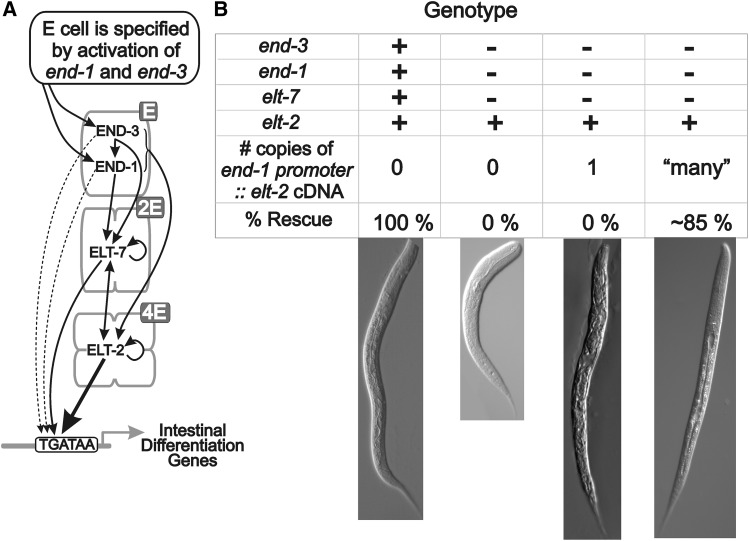
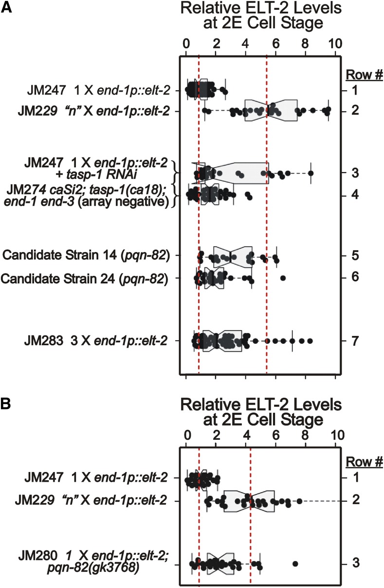
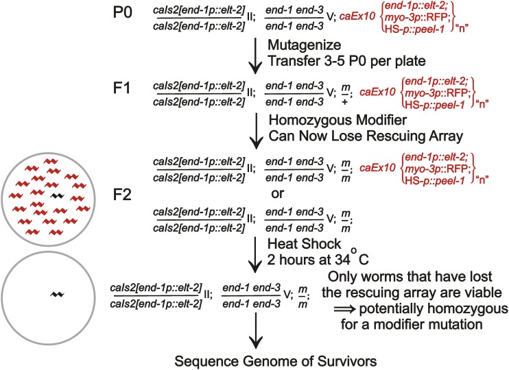
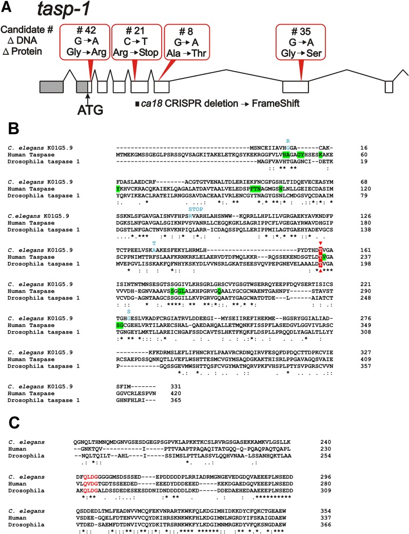
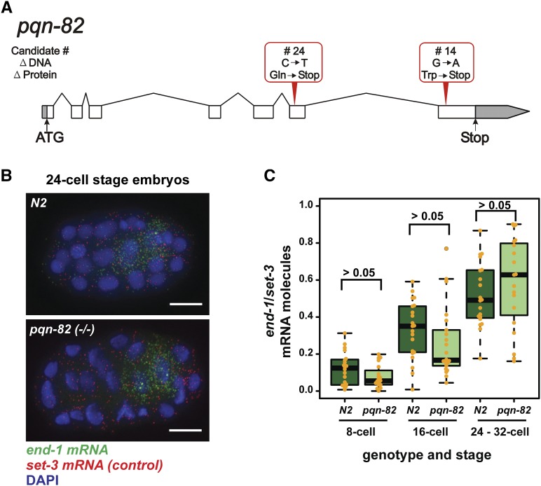
The ELT-2 GATA factor normally functions in differentiation of the C. elegans endoderm, downstream of endoderm specification. We have previously shown that, if ELT-2 is expressed sufficiently early, it is also able to specify the endoderm and to replace all other members of the core GATA-factor transcriptional cascade (END-1, END-3, ELT-7). However, such rescue requires multiple copies (and presumably overexpression) of the end-1p::elt-2 cDNA transgene; a single copy of the transgene does not rescue. We have made this observation the basis of a genetic screen to search for genetic modifiers that allow a single copy of the end-1p::elt-2 cDNA transgene to rescue the lethality of the end-1 end-3 double mutant. We performed this screen on a strain that has a single copy insertion of the transgene in an end-1 end-3 background. These animals are kept alive by virtue of an extrachromosomal array containing multiple copies of the rescuing transgene; the extrachromosomal array also contains a toxin under heat shock control to counterselect for mutagenized survivors that have been able to lose the rescuing array. A screen of ∼14,000 mutagenized haploid genomes produced 17 independent surviving strains. Whole genome sequencing was performed to identify genes that incurred independent mutations in more than one surviving strain. The C. elegans gene tasp-1 was mutated in four independent strains. tasp-1 encodes the C. elegans homolog of Taspase, a threonine-aspartic acid protease that has been found, in both mammals and insects, to cleave several proteins involved in transcription, in particular MLL1/trithorax and TFIIA. A second gene, pqn-82, was mutated in two independent strains and encodes a glutamine-asparagine rich protein. tasp-1 and pqn-82 were verified as loss-of-function modifiers of the end-1p::elt-2 transgene by RNAi and by CRISPR/Cas9-induced mutations. In both cases, gene loss leads to modest increases in the level of ELT-2 protein in the early endoderm although ELT-2 levels do not strictly correlate with rescue. We suggest that tasp-1 and pqn-82 represent a class of genes acting in the early embryo to modulate levels of critical transcription factors or to modulate the responsiveness of critical target genes. The screen's design, rescuing lethality with an extrachromosomal transgene followed by counterselection, has a background survival rate of <10-4 without mutagenesis and should be readily adapted to the general problem of identifying suppressors of C. elegans lethal mutations.

Keywords: C. elegans; ELT-2 GATA factor; Mutant Screen Report; endoderm specification; intestine; pqn-82; tasp-1; taspase.


Figures

Figure 1
Figure 1
The Waddington developmental landscape (adapted from Figure 4, Chapter 2 of (Waddington 1957)) provides a metaphor for the developmental decisions made during embryonic development. Waddington envisaged the shape of the landscape to be determined by embryonic genes that could, in principle, change the contours of the landscape, such that valleys/trajectories could be shallow and sensitive to perturbation (left) or steep and more robust (right).
Figure 2
Figure 2
A. The zygotic cascade of GATA-type transcription factors that first specify (END-1 and END-3) and then differentiate (ELT-7 and ELT-2) the C. elegans endoderm lineage. Redrawn from Wiesenfahrt et al. (2016). B. Results supporting the rationale for the present screen (Wiesenfahrt et al. 2016). 100% of wildtype animals (elt-7(+) end-1(+) end-3(+); elt-2(+)) survive. None of elt-7(-) end-1(-) end-3(-); elt-2(+) survive (see also Owraghi et al. (2009)) unless they are transgenic for multiple copies of an end-1p::elt-2 cDNA transgene. They do not survive if the transgene is present as a single copy.
Figure 3
Figure 3
Levels of immunologically detectable ELT-2 protein measured at the 2E cell stage in embryos containing either single or multiple copies of an end-1p::elt-2 transgene, with or without candidate mutations or RNAi. Each black circle corresponds to an individual embryo; the whiskers encompass all data points not judged to be outliers; the box represents the interquartile range (i.e., 25 to 75% of the data); the notch represents ∼1.6(interquartile range)/(number of observations)1/2, such that the medians of two data sets are judged to be significantly different from each other if the corresponding notches do not overlap (McGill et al. 1978). Figures 3A and 3B represent independent experiments. Fluorescent intensities are normalized to the average intensity measured with JM247 (single copy of the end-1p::elt-2 transgene) in each experiment. Dashed red lines correspond to mean (normalized) ELT-2 levels measured for strain JM247 (single copy end-1p::elt-2) or strain JM229 (multiple integrated copies of end-1p::elt-2).
Figure 4
Figure 4
Outline of the genetic screen to identify modifiers of the single copy end-1p::elt-2 cDNA transgene. A more detailed description of the rationale of each step is provided in the main text.
Figure 5
Figure 5
A. Four independent mutations identified in tasp-1 are shown above the gene. The CRISPR mutation (ca18) introduced into the gene is also shown (below). B. Alignment (Clustal Omega performed at www.ebi.ac.uk with default settings) of the protein sequences from C. elegans tasp-1, human Taspase1 and Drosophila taspase 1. The Threonine residue highlighted in red depicts the active site nucleophile, which lies immediately downstream of the aspartic acid residue in the autocleavage site. Residues highlighted in green on the sequence of human Taspase1 have been identified as part of the active site and are not found in asparaginases (Khan et al. 2005). Changes in the amino acid sequence resulting from the four candidate mutations identified in the current screen are shown in blue. C. Alignment (Clustal Omega) of the C-terminal portions of TFIIA, showing that the taspase cleavage site known in humans and Drosophila is also conserved in C. elegans (highlighted).
Figure 6
Figure 6
A. The two independent mutations identified in the structural gene of pqn-82 are shown above the gene. The genomic coordinates for the altered base pairs are 1,851,047 for Candidate # 24 and 1,852,218 for Candidate # 14. B. end-1 mRNA (green) in wild-type (N2) and pqn-82 embryos as imaged by single molecule fluorescence in situ hybridization (smFISH). Ubiquitously expressed set-3 mRNA transcripts (red) were co-hybridized as a control, and nuclei were stained with DAPI. Scale bar is 10 μm. C. Single molecules of end-1 and set-3 mRNA that were imaged by smFISH (B) were quantitated at three stages of embryonic development. Levels of set-3 transcripts stayed consistent across stages (mean of 444 molecules/embryo) but levels of end-1 transcripts increased (means of 47, 109, 214 molecules/embryo at 8-cell, 16-cell, and 32-cell stages). However, the relative amount of end-1 mRNA in N2 and pqn-82(−/−) embryos was not significantly different for each stage-specific, pair-wise comparison as calculated by Student’s two-tailed t-test (20 embryos per stage and genotype). Upper boxplot whiskers represent the lesser of either the greatest value point or the upper quartile plus 1.5 times the interquartile range; lower whiskers represent the reverse. The data derive from one of two independent replicates, both of which reach the same conclusions.

Similar articles

Cited by

References

    1. Arribere J. A., Bell R. T., Fu B. X., Artiles K. L., Hartman P. S., et al. , 2014. Efficient marker-free recovery of custom genetic modifications with CRISPR/Cas9 in Caenorhabditis elegans. Genetics 198(3): 837–846. 10.1534/genetics.114.169730 - DOI - PMC - PubMed
    1. Blackwell T. K., Walker A. K., 2006. Transcription mechanisms. WormBook 5: 1–16. - PMC - PubMed
    1. Boeck M. E., Boyle T., Bao Z., Murray J., Mericle B., et al. , 2011. Specific roles for the GATA transcription factors end-1 and end-3 during C. elegans E-lineage development. Dev. Biol. 358(2): 345–355. 10.1016/j.ydbio.2011.08.002 - DOI - PMC - PubMed
    1. Bowerman B., Draper B. W., Mello C. C., Priess J. R., 1993. The maternal gene skn-1 encodes a protein that is distributed unequally in early C. elegans embryos. Cell 74(3): 443–452. 10.1016/0092-8674(93)80046-H - DOI - PubMed
    1. Bowerman B., Eaton B. A., Priess J. R., 1992. skn-1, a maternally expressed gene required to specify the fate of ventral blastomeres in the early C. elegans embryo. Cell 68(6): 1061–1075. 10.1016/0092-8674(92)90078-Q - DOI - PubMed

Publication types

MeSH terms

Substances

LinkOut - more resources