. 2018 May 4;8(5):1807-1816.
doi: 10.1534/g3.118.200172.

Vps74 Connects the Golgi Apparatus and Telomeres in Saccharomyces cerevisiae

Affiliations

Vps74 Connects the Golgi Apparatus and Telomeres in Saccharomyces cerevisiae

Joana Rodrigues et al. G3 (Bethesda). .

Abstract

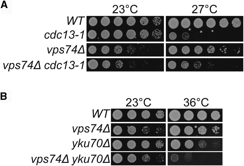
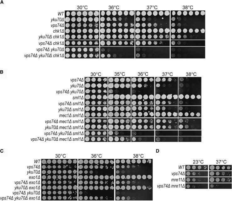
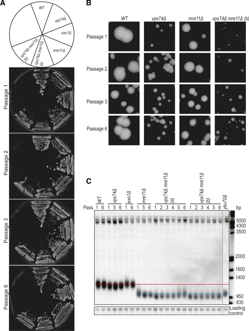
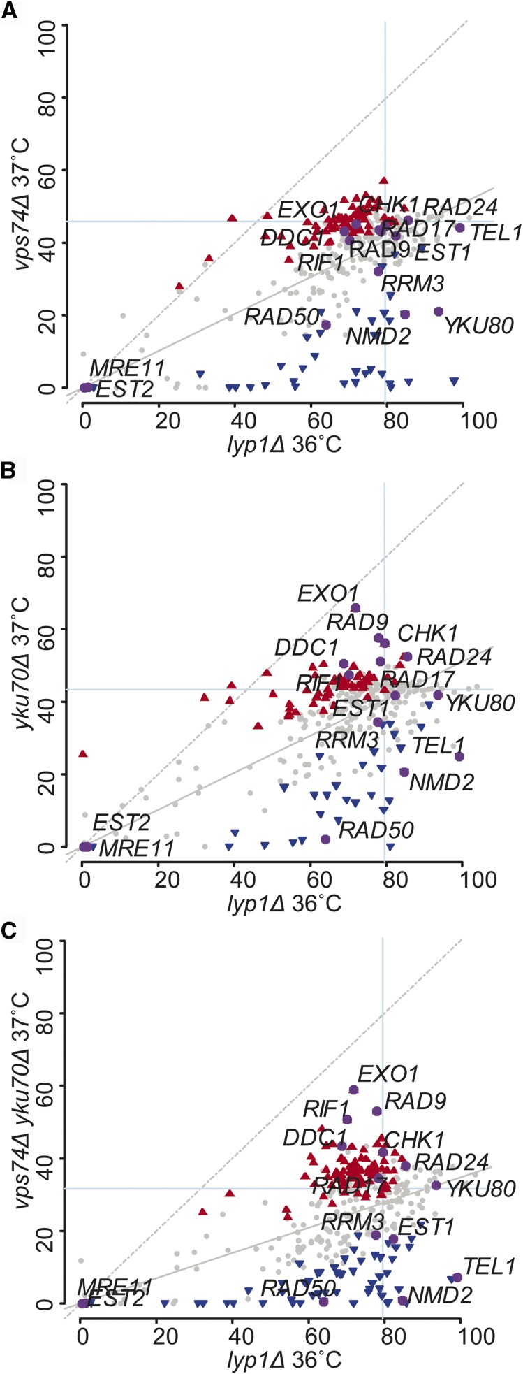
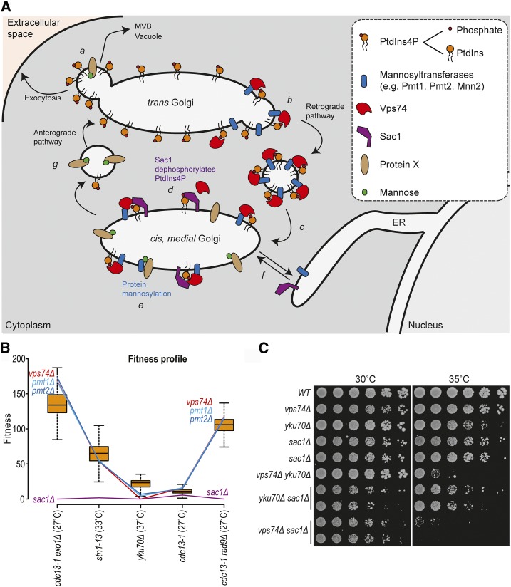
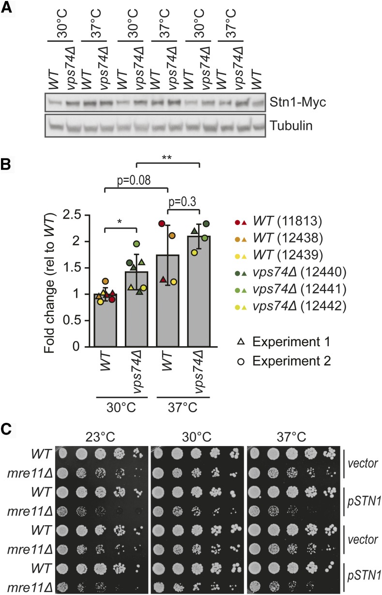
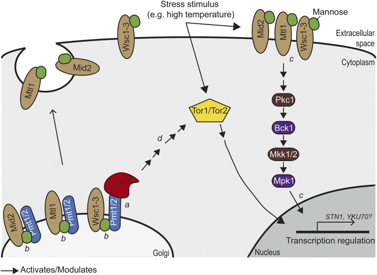
In mammalian cell culture, the Golgi apparatus fragment upon DNA damage. GOLPH3, a Golgi component, is a phosphorylation target of DNA-PK after DNA damage and contributes to Golgi fragmentation. The function of the yeast (Saccharomyces cerevisiae) ortholog of GOLPH3, Vps74, in the DNA damage response has been little studied, although genome-wide screens suggested a role at telomeres. In this study we investigated the role of Vps74 at telomeres and in the DNA damage response. We show that Vps74 decreases the fitness of telomere defective cdc13-1 cells and contributes to the fitness of yku70Δ cells. Importantly, loss of Vps74 in yku70Δ cells exacerbates the temperature dependent growth defects of these cells in a Chk1 and Mec1-dependent manner. Furthermore, Exo1 reduces the fitness of vps74Δ yku70Δ cells suggesting that ssDNA contributes to the fitness defects of vps74Δ yku70Δ cells. Systematic genetic interaction analysis of vps74Δ, yku70Δ and yku70Δ vps74Δ cells suggests that vps74Δ causes a milder but similar defect to that seen in yku70Δ cells. vps74Δ cells have slightly shorter telomeres and loss of VPS74 in yku70Δ or mre11Δ cells further shortens the telomeres of these cells. Interestingly, loss of Vps74 leads to increased levels of Stn1, a partner of Cdc13 in the CST telomere capping complex. Overexpression of Stn1 was previously shown to cause telomere shortening, suppression of cdc13-1 and enhancement of yku70Δ growth defects, suggesting that increased levels of Stn1 may be the route by which Vps74 affects telomere function. These results establish Vps74 as a novel regulator of telomere biology.

Keywords: Golgi; QFA; Saccharomyces cerevisiae; Stn1; Vps74; telomere.


Figures

Figure 1
Figure 1
VPS74 affects the fitness of telomere defective cells. (A and B) Serial dilutions of saturated overnight cultures, grown at 23°C, were spotted onto YEPD plates and incubated for 2 days at the indicated temperatures. All strains are in the W303 genetic background and at each temperature were grown on a single plate but images have been cut and pasted to allow better comparisons.
Figure 2
Figure 2
CHK1, MEC1, EXO1 and MRE11 affect the fitness of vps74Δ yku70Δ and vps74Δ cells at high temperatures. Spot test assays as described in Figure 1.
Figure 3
Figure 3
Loss of VPS74 leads to telomere shortening in WT and mre11Δ cells. (A) Passage tests performed at 30°C. Cells were allowed to grow for 2 days before pictures were taken and cells passaged. (B) Zoom in of the colonies in A. (C) Cells from the plates in A were inoculated in liquid YEPD, grown until saturation and DNA was extracted. The DNA was analyzed by Southern blot with a telomere probe (Y’+TG). Horizontal red line represents the WT telomere length and the blue line is roughly the telomere length of mre11Δ cells. Vertical dashed line indicates where the gel picture was cut for presentation purposes.
Figure 4
Figure 4
Systematic analysis of the effects of 358 gene deletions in the fitness of vps74Δ, yku70Δ and yku70Δ vps74Δ cells. Part of the yeast genome knock out collection (358 strains, Table S3) was crossed with vps74Δ (A), yku70Δ (B), vps74Δ yku70Δ (C) or lyp1Δ (A, B and C) mutations. Double or triple mutants were then grown on solid agar plates and the fitness was measured at 37°C (or 36°C for lyp1Δ). Fitness is measured as Maximum Doubling Rate × Maximum Doubling Potential (MDR × MDP, units are doublings squared per day, d2/day), as previously described (addinall et al. 2011). Each dot indicates the effect of a gene deletion (yfgΔ) on the fitness of vps74Δ (A), yku70Δ (B), vps74Δ yku70Δ (C) or lyp1Δ (A, B and C). Gray dots indicate the deletions that did not significantly alter the fitness of the mutants in the y axis relative to the x axis (lyp1Δ). Blue inverted triangles represent gene deletions that are enhancers of the vps74Δ (A), yku70Δ (B) or vps74Δ yku70Δ (C), red triangles are suppressors and the purple dots represent the fitness of the deletion of 15 telomere related genes. Figures showing vps74Δ screens vs. yku70Δ screens, vps74Δ yku70Δ screens vs. yku70Δ screens and vps74Δ yku70Δ screens vs. vps74Δ screens can be found in Figure S2B, C and D, respectively.
Figure 5
Figure 5
vps74Δ, pmt1Δ and pmt2Δ show similar genetic interactions with mutations affecting telomeres. (A) Cartoon showing the Vps74 function in protein sorting within the Golgi apparatus (schmitz et al. 2008; wood et al. 2012). Complex sphingolipids are preferentially packaged with secretory cargo into anterograde-directed transport vesicles that end up in being exocytosed, in multivesicular bodies (MVB) or the vacuole (a). In the trans Golgi, Vps74 recognizes PtdIns4P and mannosyltransferases, sorting them into COPI-coated retrograde vesicles (b). As a consequence of co-packaging PtdIns4P with Golgi protein residents into retrograde vesicles, PtdIns4P is delivered to the medial cisternae (c). In the medial/cis Golgi, Vps74 promotes Sac1-dependent PtdIns4P dephosphorylation (d) and protein mannosylation (e). Sac1 and mannosyltransferases cycle between the endoplasmic reticulum (ER) and the Golgi apparatus (f). Similarly, mannosylated proteins could follow the anterograde pathway (g, a) or eventually return to ER (f). Cartoon and legend adapted from (wood et al. 2012). (B) Fitness profiles of vps74Δ, sac1Δ, pmt1Δ and pmt2Δ in combination with several mutations affecting telomere biology (x axis) (holstein et al. 2017). Box plots show 50% range, the whiskers represent 1.5-fold the 50% range from the box, and the horizontal black line is the median fitness. (C) Spot test assays as described in Figure 1.
Figure 6
Figure 6
Increased temperature and loss of Vps74 leads to increased Stn1-Myc levels. Three independent WT and vps74Δ strains carrying a Stn1-Myc construct (strains numbers indicated), were cultured overnight at 30°C to saturation. Each culture was diluted 1:100, cultured for 2h at 30°C and then divided into two cultures that were further incubated at 30°C or 37°C for 4h. (A) Proteins were extracted using TCA and a western blot was performed first against Myc and then against tubulin. (B) Quantification of A and Figure S6 using Image J. Stn1-Myc intensity normalized for tubulin levels is shown. Values are presented as fold change relative to WT at 30°C. At 30°C, three independent strains were analyzed in two independent experiments (n = 6), while at 37° two independent strains were analyzed once while a third independent strain was analyzed twice (independent experiments, n = 4). The mean is indicated and the error bars indicate the standard deviation. Statistical analyses used the two-tailed t-Test assuming unequal variance (*P < 0.05 and **P < 0.01) performed with SigmaPlot (version 11). (C) WT and mre11Δ cells were transformed with a 2 µm plasmid carrying STN1 or a vector plasmid (plasmids described in Table S4). Six independent (WT and mre11Δ) transformants carrying either the STN1 plasmid or the vector, were cultivated in selective media (-LEU, lacking leucine) until saturation and spot tests were performed as described in Figure 1 (–LEU plates). Pictures were taken after 3 days of incubation. Two representative strains of each genotype carrying either of the plasmids are shown.
Figure 7
Figure 7
Speculative model for how Vps74 regulates stress responses through the Tor1/2 and Mtl1 pathways. Model for the possible role of Vps74 regulating gene transcription and protein levels in response to stress. (a) We suggest that Vps74 is important for the function of the mannosyltransferases Pmt1 and Pmt2 by helping recruit them to Golgi. (b) Pmt1 and Pmt2 mediate O-mannosylation of the transmembrane receptors Wsc1-3, Mid2 and Mtl1 (ketela et al. 1999; philip and levin 2001; lommel et al. 2004). Mannosylated Wsc1-3, Mid2 and Mtl1 are delivered to cytoplasmic membrane. (c) O-mannosylation of transmembrane receptors is important for proper activation of the Pkc1-Bck1-Mkk1/2-Mpk1 stress response cascade pathway Mtl1 (ketela et al. 1999; philip and levin 2001; lommel et al. 2004). The Pkc1-Bck1-Mkk1/2-Mpk1 cascade pathway promotes transcriptional changes and Mpk1 was shown to promote telomere silencing, transcription modulation and proteasome homeostasis (ai et al. 2002; jendretzki et al. 2011; lee et al. 2013; rousseau and bertolotti 2016). (d) Data from mammalian cells showed that GOLPH3 (Vps74 human ortholog) promotes mTor (Tor1/2 in yeast) activation (ai et al. 2002; scott et al. 2009; jendretzki et al. 2011; lee et al. 2013; rousseau and bertolotti 2016). The Tor pathway modulates gene transcription and protein translation in response to nutrient availability and cellular stresses.

Similar articles

Cited by

References

    1. Adams A., Gottschling D. E., Kaiser C. A., Stearns T., 1997. Methods in Yeast Genetics, Cold Spring Harbor Laboratory Press, New York.
    1. Addinall S. G., Holstein E. M., Lawless C., Yu M., Chapman K., et al. , 2011. Quantitative Fitness Analysis Shows That NMD Proteins and Many Other Protein Complexes Suppress or Enhance Distinct Telomere Cap Defects. PLoS Genet. 7: e1001362 10.1371/journal.pgen.1001362 - DOI - PMC - PubMed
    1. Ai W., Bertram P. G., Tsang C. K., Chan T. F., Zheng X. F., 2002. Regulation of subtelomeric silencing during stress response. Mol. Cell 10(6): 1295–1305. 10.1016/S1097-2765(02)00695-0 - DOI - PubMed
    1. Brameier M., Krings A., MacCallum R. M., 2007. NucPred–predicting nuclear localization of proteins. Bioinformatics 23(9): 1159–1160. 10.1093/bioinformatics/btm066 - DOI - PubMed
    1. Cai Y., Deng Y., Horenkamp F., Reinisch K. M., Burd C. G., 2014. Sac1-Vps74 structure reveals a mechanism to terminate phosphoinositide signaling in the Golgi apparatus. J. Cell Biol. 206(4): 485–491. 10.1083/jcb.201404041 - DOI - PMC - PubMed

Publication types

Substances