. 2018 May;49(5):1232-1240.
doi: 10.1161/STROKEAHA.117.020356. Epub 2018 Mar 28.

Thalidomide Reduces Hemorrhage of Brain Arteriovenous Malformations in a Mouse Model

Affiliations

Thalidomide Reduces Hemorrhage of Brain Arteriovenous Malformations in a Mouse Model

Wan Zhu et al. Stroke. 2018 May.

Abstract

Background and purpose: Brain arteriovenous malformation (bAVM) is an important risk factor for intracranial hemorrhage. Current treatments for bAVM are all associated with considerable risks. There is no safe method to prevent bAVM hemorrhage. Thalidomide reduces nose bleeding in patients with hereditary hemorrhagic telangiectasia, an inherited disorder characterized by vascular malformations. In this study, we tested whether thalidomide and its less toxic analog, lenalidomide, reduce bAVM hemorrhage using a mouse model.

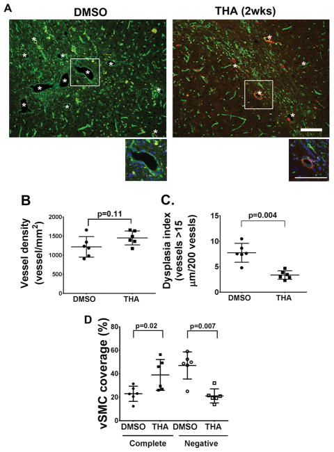
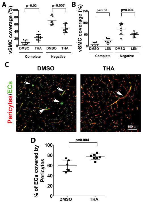
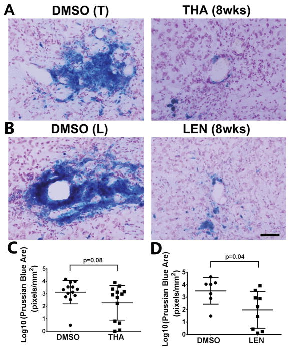
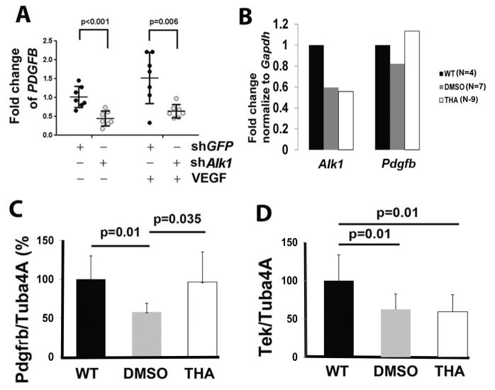
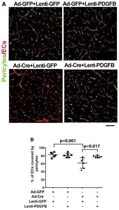
Methods: bAVMs were induced through induction of brain focal activin-like kinase 1 (Alk1, an AVM causative gene) gene deletion and angiogenesis in adult Alk1-floxed mice. Thalidomide was injected intraperitoneally twice per week for 6 weeks, starting either 2 or 8 weeks after AVM induction. Lenalidomide was injected intraperitoneally daily starting 8 weeks after AVM induction for 6 weeks. Brain samples were collected at the end of the treatments for morphology, mRNA, and protein analyses. The influence of Alk1 downregulation on PDGFB (platelet-derived growth factor B) expression was also studied on cultured human brain microvascular endothelial cells. The effect of PDGFB in mural cell recruitment in bAVM was explored by injection of a PDGFB overexpressing lentiviral vector to the mouse brain.

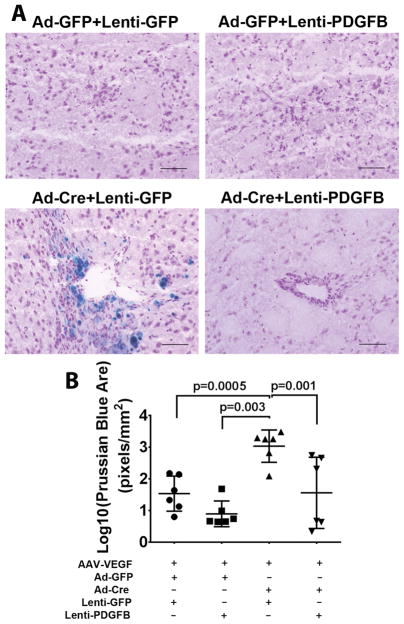
Results: Thalidomide or lenalidomide treatment reduced the number of dysplastic vessels and hemorrhage and increased mural cell (vascular smooth muscle cells and pericytes) coverage in the bAVM lesion. Thalidomide reduced the burden of CD68+ cells and the expression of inflammatory cytokines in the bAVM lesions. PDGFB expression was reduced in ALK1-knockdown human brain microvascular endothelial cells and in mouse bAVM lesion. Thalidomide increased Pdgfb expression in bAVM lesion. Overexpression of PDGFB mimicked the effect of thalidomide.

Conclusions: Thalidomide and lenalidomide improve mural cell coverage of bAVM vessels and reduce bAVM hemorrhage, which is likely through upregulation of Pdgfb expression.

Keywords: arteriovenous malformation; brain; endothelial cells; hemorrhage; thalidomide.


Conflict of interest statement

Conflict of Interests:

The authors have declared that no conflict of interest exists.

Figures

Figure 1
Figure 1. Thalidomide inhibits bAVM development
A. Representative images of brain sections. Vessels were stained by lectin (green). Vascular smooth muscles were stained with an antibody against α-smooth muscle actin (red). *: indicates dysplastic vessels. The enlarged images of white rectangle areas are shown below the pictures. Scale bars: 100 μm. B. Quantification of vessel density. P=0.11, by t-test analysis. C. Dysplasia index (numbers of vessels that are larger than 15 μm in diameter per 200 vessels). P=0.004, by t-test analysis. D. Quantification of vSMC coverage. The data are the percentage of dysplastic vessels that were covered (Complete) or not covered (Negative) by vSMCs. THA (2wks): mice received thalidomide treatment starting 2 weeks after model induction. P values were determined by one-way ANOVA followed by Sidak’s multiple comparisons. N=6 for all analyses.
Figure 2
Figure 2. Thalidomide and lenalidomide increase mural cell-coverage in established bAVMs
A & B. Quantification of vSMC (α-smooth muscle actin positive cells)-coverage. The data are the percentage of dysplastic vessels that were covered (complete) or not covered (Negative) vSMCs. N=8 for thalidomide (THA) treated group and its DMSO control. N=8 for lenalidomide (LEN) treated group and N=7 for its DMSO control. P values were determined by one-way ANOVA followed by Sidak’s multiple comparisons. C. Representative confocal images of brain sections stained with antibodies specific to endothelial cells (CD31, green) and pericytes (CD13, red). Arrows indicate pericyte negative vessel segments. Scale bars: 100 μm. D. Quantification of the percentage of endothelial cells (ECs) covered by pericytes. N=7 for thalidomide (THA) treated group and N=6 for its DMSO control. P=0.004, which was determined by one-way ANOVA followed by Sidak’s multiple comparisons. 8wks: treatments starting 8 weeks after model induction.
Figure 3
Figure 3. Thalidomide and lenalidomide reduces hemorrhage in the established bAVM
A & B. Representative images of Prussian blue stained sections. Scale bar: 100 μm. C & D. Quantification of Prussian blue-positive area. Log 10: the data were 10 log conversed. THA (8wks): mice received thalidomide treatment starting 8 weeks after model induction. LEN (8wks): mice received lenalidomide treatment starting 8 weeks after model induction. DMSO (T): control for thalidomide treated-group; DMSO (L): control for lenalidomide treated-group. N=13 for thalidomide group and its DMSO control. N=9 for lenalidomide group and N=7 for its DMSO control. P values were determined by t-test analysis.
Figure 4
Figure 4. Thalidomide treatment upregulated Pdgfb and Pdgfrβ expression
A. PDGFB expression was reduced in ALK1 knockdown HBMECs. The Y axis is fold changes compared to the mean of Lenti-GFP (shGFP) treated controls without VEGF treatment. N=7. P values were determined by one-way ANOVA analysis followed by Sidak’s multiple comparisons. B. Quantification of Alk1 and Pdgfb expression in bAVM microvessels. In order to obtained enough RNA for the analysis. microvessels from mice in same group were pooled: 4 wild type (WT) mice, 7 DMSO treated mice and 9 thalidomide (THA) treated mice. C & D. Quantification of western blot results. Tuba4a was used as an internal control for normalization of protein load. Data was shown by percentage (%) normalized to the WT group. N=6. P values were determined by one-way ANOVA followed by Sidak’s multiple comparisons.
Figure 5
Figure 5. Overexpression of PDGFB increases pericyte covered bAVM vessels
A. Representative confocal images of brain sections stained with antibodies specific to CD31 (an endothelial cell-marker, red) and CD13 (a pericyte-marker, green). Scale bars: 100 μm. B. Quantification of the percentage of endothelial cells (ECs) covered by pericypes. All mice were Alk12f/2f mice and were treated with AAV-VEGF to induce brain angiogenesis. Ad-GFP+Lenti-GFP: Controls for normal angiogenesis; Ad-GFP+Lenti-PDGFB: Overexpressing PDGFB in angiogenic brain; Ad-Cre+Lenti-GFP: Untreated bAVM; Ad-Cre+Lenti-PDGFB; Overexpression of PDGFB in bAVM. N=6. P values were determined by one-way ANOVA followed by Sidak’s multiple comparisons.
Figure 6
Figure 6. Overexpression of PDGFB reduced hemorrhage in the bAVM lesions
A. Representative images of Prussian blue stained sections. Scale bar: 100 μm. B. Quantification of Prussian blue positive area. The data are 10 log conversed (Log 10). All mice are treated with AAV-VEGF to induce brain angiogenesis. Ad-GFP+Lenti-GFP: Controls for normal angiogenesis; Ad-GFP+Lenti-PDGFB: Overexpressing PDGFB in angiogenic brain; Ad-Cre+Lenti-GFP: Untreated bAVM; Ad-Cre+Lenti-PDGFB; Overexpression of PDGFB in bAVM. N=6. P values were determined by one-way ANOVA followed by Sidak’s multiple comparisons.

References

    1. Kim H, Su H, Weinsheimer S, Pawlikowska L, Young WL. Brain arteriovenous malformation pathogenesis: A response-to-injury paradigm. Acta Neurochir Suppl. 2011;111:83–92. - PMC - PubMed
    1. Mohr JP, Parides MK, Stapf C, Moquete E, Moy CS, Overbey JR, et al. Medical management with or without interventional therapy for unruptured brain arteriovenous malformations (aruba): A multicentre, non-blinded, randomised trial. Lancet. 2014;383:614–621. - PMC - PubMed
    1. Sanders RD, Avidan MS. Postoperative cognitive trajectories in adults: The role of inflammatory processes. Anesthesiology. 2013;118:484–486. - PubMed
    1. Walker EJ, Su H, Shen F, Choi EJ, Oh SP, Chen G, et al. Arteriovenous malformation in the adult mouse brain resembling the human disease. Ann Neurol. 2011;69:954–962. - PMC - PubMed
    1. Chen W, Guo Y, Walker EJ, Shen F, Jun K, Oh SP, et al. Reduced mural cell coverage and impaired vessel integrity after angiogenic stimulation in the alk1-deficient brain. Arterioscler Thromb Vasc Biol. 2013;33:305–310. - PMC - PubMed

Publication types

MeSH terms