. 2018 Mar 28;9(1):1251.
doi: 10.1038/s41467-018-03632-y.

Structural characterization of a highly-potent V3-glycan broadly neutralizing antibody bound to natively-glycosylated HIV-1 envelope

Affiliations

Structural characterization of a highly-potent V3-glycan broadly neutralizing antibody bound to natively-glycosylated HIV-1 envelope

Christopher O Barnes et al. Nat Commun. .

Abstract

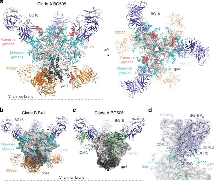
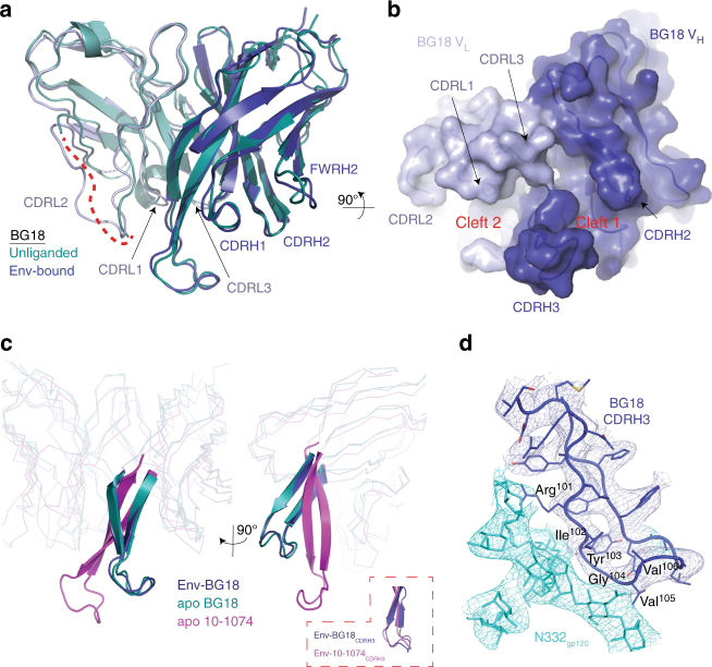
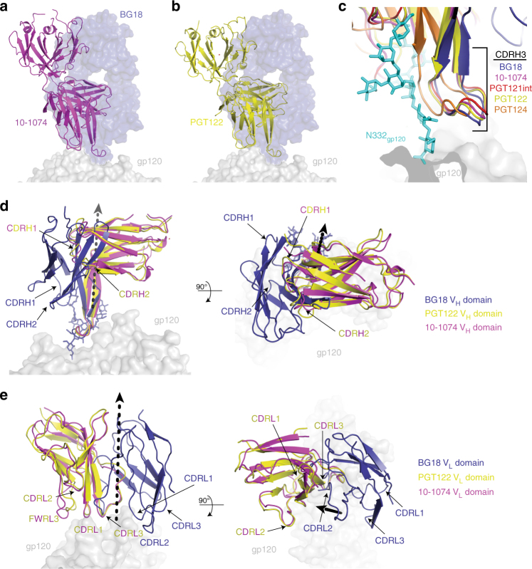
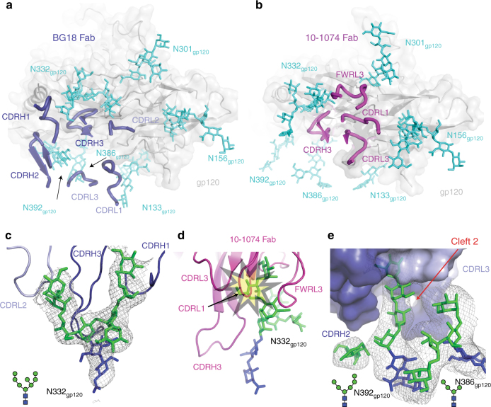
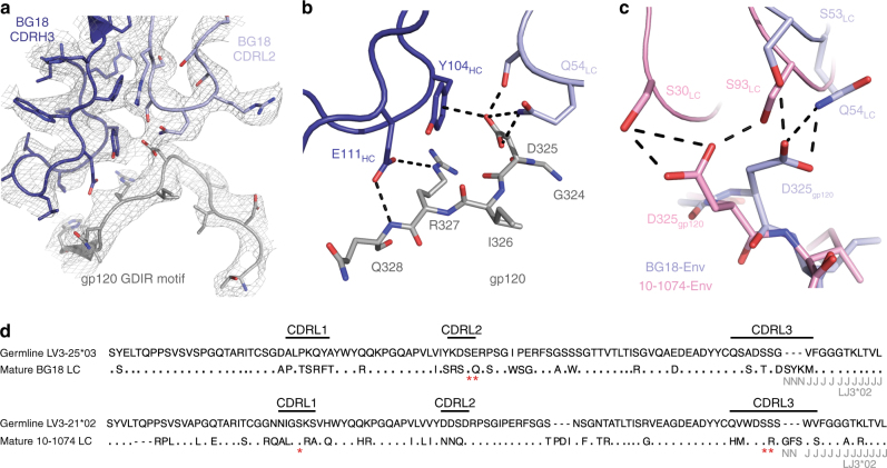
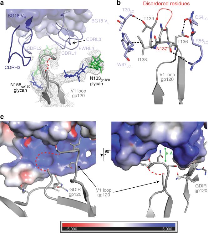
Broadly neutralizing antibodies (bNAbs) isolated from HIV-1-infected individuals inform HIV-1 vaccine design efforts. Developing bNAbs with increased efficacy requires understanding how antibodies interact with the native oligomannose and complex-type N-glycan shield that hides most protein epitopes on HIV-1 envelope (Env). Here we present crystal structures, including a 3.8-Å X-ray free electron laser dataset, of natively glycosylated Env trimers complexed with BG18, the most potent V3/N332gp120 glycan-targeting bNAb reported to date. Our structures show conserved contacts mediated by common D gene-encoded residues with the N332gp120 glycan and the gp120 GDIR peptide motif, but a distinct Env-binding orientation relative to PGT121/10-1074 bNAbs. BG18's binding orientation provides additional contacts with N392gp120 and N386gp120 glycans near the V3-loop base and engages protein components of the V1-loop. The BG18-natively-glycosylated Env structures facilitate understanding of bNAb-glycan interactions critical for using V3/N332gp120 bNAbs therapeutically and targeting their epitope for immunogen design.


Conflict of interest statement

The authors declare no competing interests.

Figures

Fig. 1
Fig. 1
Crystal structures of natively glycosylated HIV-1 Env trimers complexed with BG18. a Cartoon representation of the 3.8 Å structure of BG505 Env (gp120, light gray; gp41, dark gray) in complex with BG18 (blue) and 35O22 (orange) Fabs. Ordered, native high-mannose glycans (cyan) are represented as sticks, and complex-type glycans (salmon) are shown as spheres. b 4.9 Å structure of B41 Env bound to BG18 (blue) and 35O22 (orange). Glycans shown as cyan spheres. c 6.7 Å structure of BG505 Env bound to BG18 (blue) and IOMA (green) Fabs. Glycans were not modeled due to the limited resolution. d Close-up of the BG18 binding site (VH in dark blue, VL in light blue superimposed on the final 2FoFcalc electron density map contoured at 1.2σ) on the surface of gp120 (gray) from the BG18-BG505-35O22 structure. Ordered glycans (cyan) near the BG18 binding site are represented as sticks
Fig. 2
Fig. 2
Comparison of apo- and Env-bound BG18 variable domains. a Superposition of VH-VL domains (230 Cα atoms) of unliganded BG18 (deep teal; PDB 5UD9 (http://dx.doi.org10.2210/pdb5UD9/pdb)) with BG18 in the BG18-BG505 structure (blue) showed conservation of the BG18 antigen-binding site and ordering of CDRL2 in the BG505-bound structure (dashed red line represents disordered CDRL2 in unliganded BG18). b Surface representation of BG18 in the BG505-bound structure showed conservation of BG18 clefts, including the cleft between CDRH3 and CDRL1/3 loops observed in the BG18 apo structure. c Ribbon and cartoon representation of the overlay between BG18 (aligned on the Fab VH domains) in the BG18-BG505-35O22 complex (blue), unliganded BG18 (deep teal), and unliganded 10-1074 (magenta; PDB 4FQ2 (http://dx.doi.org10.2210/pdb4FQ2/pdb)). CDRH3 loops for the three structures are represented as cartoons. Inset: overlay of BG18-BG505 and 10-1074-BG505 (PDB 5T3Z (http://dx.doi.org10.2210/pdb5T3Z/pdb)) CDRH3 loops. d Cartoon and stick representation of BG18 CDRH3 recognition of the N332gp120 glycan. BG18 residues comprising a conserved structural motif (R-I-Y-G-V/I-I) are labeled. Electron density contoured at 1σ from 2FobsFcalc composite annealed omit map calculated with phases from models with the N332gp120 glycan and BG18 CDRH3 coordinates omitted to reduce potential phase bias (cyan and blue mesh)
Fig. 3
Fig. 3
Distinct orientation on HIV-1 Env of BG18 compared to other V3/N332gp120 bNAbs. BG18 Fab (blue, surface) orientation on gp120 (gray, surface) overlaid with 10-1074 (a, magenta; PDB 5T3Z (http://dx.doi.org10.2210/pdb5T3Z/pdb)) and PGT122 (b, yellow; 5FYL (http://dx.doi.org10.2210/pdb5T3Z/pdb)) and PGT122 (b, yellow; 5FYL (http://dx.doi.org10.2210/pdb5FYL/pdb)) Fabs. c Overlay of CDRH3 loop of BG18 (blue), 10-1074 (magenta), PGT122 (yellow), PGT121 precursor (red; PDB 5CEZ (http://dx.doi.org10.2210/pdb5CEZ/pdb)), and PGT124 (orange; PDB 4R2G (http://dx.doi.org10.2210/pdb4R2G/pdb)) after alignment of bound gp120s (gray, surface) demonstrating a conserved structural motif recognizing the N332gp120 glycan (cyan). d, e Comparison of VH-VL domains of PGT122 (yellow) and 10-1074 (magenta) with BG18 (blue). The VH-VL domain orientation of BG18 on Env trimer is related by a 78˚ rotation and 6.8 Å translation about the indicated axis (black dashed arrow) to the 10-1074 variable domains after alignment against gp120 in the BG18-Env and 10-1074-Env structures
Fig. 4
Fig. 4
Glycan interactions with V3/N332gp120 bNAbs in structures including natively glycosylated Env. a, b Comparison of the orientations on gp120 (gray surface and cartoon) of the CDR loops from (a) BG18 (blue, ribbon) and (b) 10-1074 (magenta, ribbon) demonstrating that the BG18 variable domains are rotated clockwise about CDRH3 relative to the 10-1074 variable domains. The distinct BG18 orientation on gp120 resulted in contacts with N156gp120, N386gp120, and N392gp120 glycans (cyan, sticks) in proximity to the V3-base. Red dotted outline: Differences in the N332gp120 glycan conformation on BG505 Env bound to BG18 (a) or to 10-1074 (b). c Close-up of BG18 interaction with the N332gp120 glycan showing CDRH3, CDRH1 and CDRL2 loops at the glycan interface. d Overlay of BG18-BG505 and 10-1074–BG505 (PDB 5T3Z (http://dx.doi.org10.2210/pdb5T3Z/pdb)) structures showed that the BG18-bound N332gp120 glycan conformation would clash (yellow star) with light chain CDR loops of 10-1074 and other PGT121-like bNAbs that display nearly identical binding modes. e Surface representation of BG18 interactions with the N392gp120 and N386gp120 glycans showing the N392gp120 glycan buried (~800 Å2 total BSA) inside cleft 2 located between the CDRH3 and CDRL1/3 loops. The N386gp120 glycan associates weakly with light chain BG18 (45 Å2 total BSA), but forms branch–branch interactions with the N392gp120 glycan. c, e Electron density contoured at 1σ from 2FobsFcalc composite annealed omit maps calculated with phases from models with glycan coordinates omitted to reduce potential phase bias (gray mesh)
Fig. 5
Fig. 5
Comparison of GDIR recognition by BG18 and 10-1074. a Cartoon and stick representation of BG18 CDRH3 (dark blue), BG18 CDL2 (light blue), and gp120 (gray; residues 318-330). Electron density contoured at 1σ from 2FobsFcalc final refined electron density map with −100 Å2 B-sharpening is shown (gray mesh). b BG18 interactions with the gp120 GDIR motif (G324gp120-D325gp120-I326gp120-R327gp120) at the base of the V3-loop. In common with the PGT121-like bNAbs, CDRH3 E111HC forms a potential salt bridge with R327gp120, while CDRH3 Y104HC stacks against D325gp120. In addition, CDRL2 residues S53LC and Q54LC engage D325gp120 forming potential H-bond interactions. c Comparison of BG18 (light blue) and 10-1074 (pink; PDB 5T3Z (http://dx.doi.org10.2210/pdb5T3Z/pdb)) interactions with D325gp120 in the GDIR motif. Engagement of the carboxylate group of D325gp120 is achieved by residues in CDRL2 (BG18) or serine residues in CDRs L1 and L3 (10-1074). Potential H-bonds represented as dashed lines. d Alignment of sequences of inferred germline and mature light chains of BG18 and 10-1074. The LV3-21*02 germline V gene segment is used for both 10-1074 and PGT121-like antibodies. Red asterisks indicate residues involved in D325gp120 recognition in the GDIR motif as shown in c
Fig. 6
Fig. 6
BG18 contacts with gp120 V1-loop. a Surface and cartoon representation of BG18 VH (dark blue) and VL (light blue) loops involved in gp120 V1-loop (gray; residues 128–158, disordered residues depicted as dashed red line) interactions. Electron density contoured at 1σ from a 2FobsFcalc composite annealed omit map is shown (gray mesh). Glycans at position N133gp120 and N156gp120 are shown. b Cartoon and stick representation of residue interactions between BG18 VL domain (light blue) and the gp120 V1-loop (gray). Potential H-bonding occurs between T139gp120 and BG18 T30LC in the CDRL1 loop. In addition, BG18 W67LC in FWRL3 stacks against I138gp120. BG18 contacts with gp120 positions 138 and 139 are likely specific to Envs with V1 characteristics similar to BG505, since similar conformations were not observed in our BG18-B41 structure. H-bonds and pi-stacking are indicated by black dashed lines. Red asterisk on N137gp120 indicates a PNGS. c Electrostatic surface potentials with red indicating negative electrostatic potential and blue indicating positive electrostatic potential for BG18 shown with cartoon and stick representation of nearby gp120 elements. BG18 includes a positively charged cleft in the vicinity of the gp120 V1-loop (dashed line indicates disordered region), which may provide increased protein–protein interactions with the gp120 surface in HIV-1 strains harboring charged residues in the V1-loop

References

    1. Klein F, et al. HIV therapy by a combination of broadly neutralizing antibodies in humanized mice. Nature. 2012;492:118–122. doi: 10.1038/nature11604. - DOI - PMC - PubMed
    1. Horwitz JA, et al. HIV-1 suppression and durable control by combining single broadly neutralizing antibodies and antiretroviral drugs in humanized mice. Proc. Natl Acad. Sci. USA. 2013;110:16538–16543. doi: 10.1073/pnas.1315295110. - DOI - PMC - PubMed
    1. Shingai M, et al. Antibody-mediated immunotherapy of macaques chronically infected with SHIV suppresses viraemia. Nature. 2013;503:277–280. doi: 10.1038/nature12746. - DOI - PMC - PubMed
    1. Barouch DH, et al. Therapeutic efficacy of potent neutralizing HIV-1-specific monoclonal antibodies in SHIV-infected rhesus monkeys. Nature. 2013;503:224–228. doi: 10.1038/nature12744. - DOI - PMC - PubMed
    1. Bar KJ, et al. Effect of HIV Antibody VRC01 on Viral Rebound after Treatment Interruption. N. Engl. J. Med. 2016;375:2037–2050. doi: 10.1056/NEJMoa1608243. - DOI - PMC - PubMed

Publication types

MeSH terms