. 2018 Mar 28;8(1):5306.
doi: 10.1038/s41598-018-23714-7.

Vitamin C promotes apoptosis in breast cancer cells by increasing TRAIL expression

Affiliations

Vitamin C promotes apoptosis in breast cancer cells by increasing TRAIL expression

David W Sant et al. Sci Rep. .

Abstract

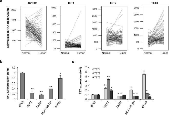
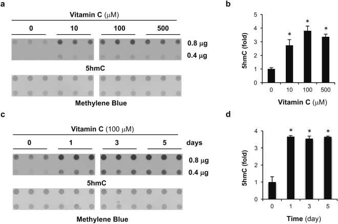
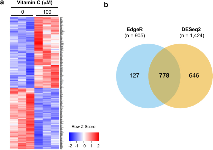
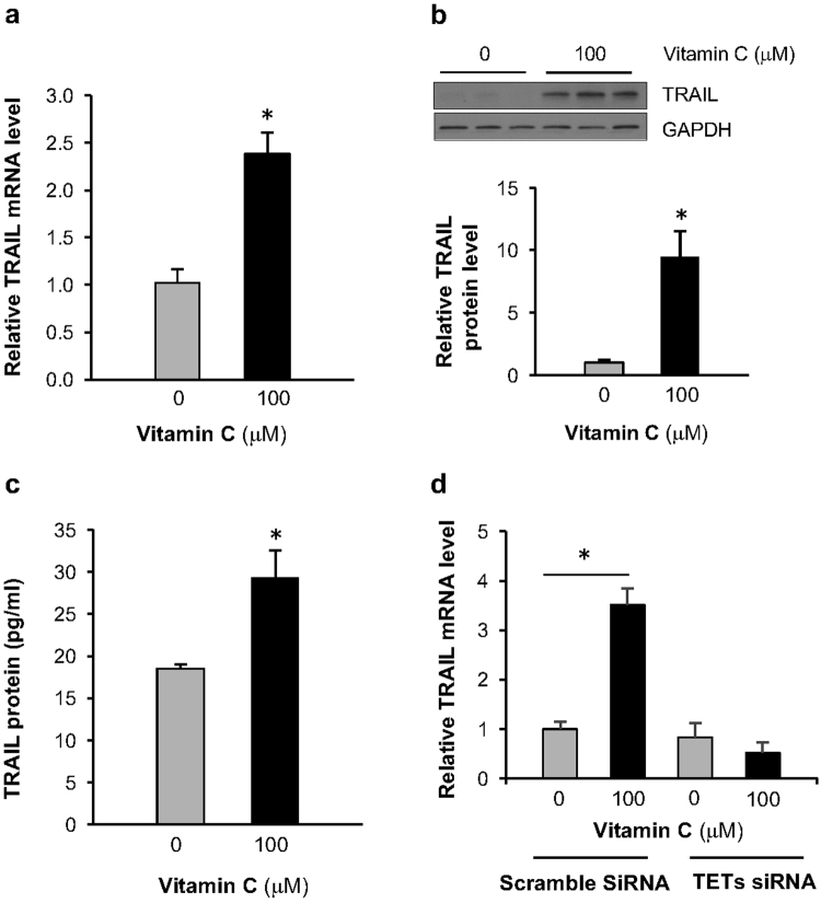
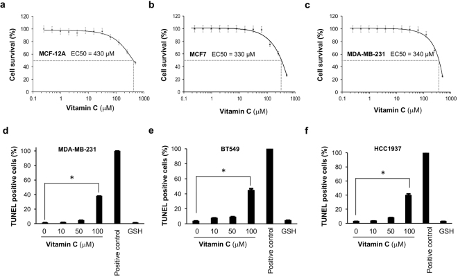
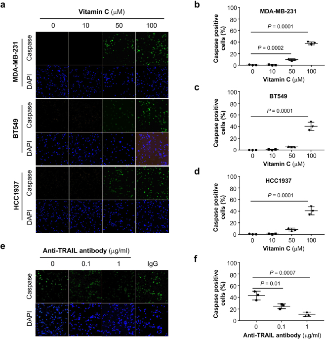
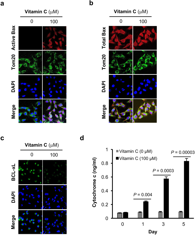
Genomic loss of 5-hydroxymethylcytosine (5hmC) accompanies malignant cellular transformation in breast cancer. Vitamin C serves as a cofactor for TET methylcytosine dioxygenases to increase 5hmC generation. Here we show that the transcription of SVCT2, a major vitamin C transporter, was decreased in human breast cancers (113 cases) compared to normal breast tissues from the same patients. A decreased SVCT2 expression was also observed in breast cancer cell lines. Treatment with vitamin C (100 μM) increased the 5hmC content in MDA-MB-231 breast cancer cells and markedly altered the transcriptome. The vitamin C treatment induced apoptosis in MDA-MB-231 cells, which was verified in two additional breast cancer cell lines. This pro-apoptotic effect of vitamin C appeared to be mediated by TRAIL, a known apoptosis inducer. Vitamin C upregulated TRAIL transcripts (2.3-fold increase) and increased TRAIL protein levels. The upregulation of TRAIL by vitamin C was largely abolished by siRNAs targeting TETs and anti-TRAIL antibody abrogated the induction of apoptosis. Furthermore, the apoptosis promoted by vitamin C was associated with Bax and caspases activation, Bcl-xL sequestration, and cytochrome c release. Taken together, these results suggest a potential role of physiological doses of vitamin C in breast cancer prevention and treatment.


Conflict of interest statement

The authors declare no competing interests.

Figures

Figure 1
Figure 1
Downregulation of SVCT2 in breast cancer. (a) RNA-seq of 113 paired normal breast tissue/breast cancer available in the TCGA dataset shows that the mRNA levels of SVCT2 are decreased in many cancer samples compared to controls (P = 2.31 × 10−19). In contrast TETs remain unchanged or increased in the majority of breast cancer cases. Each line represents an individual patient, linking the level of mRNA in the normal tissue and tumor tissue from that specific patient. (b) Quantitative RT-PCR shows that SVCT2 mRNA levels were decreased in breast cancer cell lines. (c) The mRNA level of TETs was either increased or decreased in breast cancer cell lines shown by quantitative RT-PCR.
Figure 2
Figure 2
Vitamin C treatment elevates 5hmC content in breast cancer cells. (a) The dot-blot shows that treatment with 10, 100, and 500 μM vitamin C for 24 hours increases the global content of 5hmC. (b) The semi-quantitation of the dot-blot shows that 10 μM vitamin C treatment for 24 hours increases the global content of 5hmC, supplementation to 100 μM further increases 5hmC levels, but further supplementation to 500 μM does not increase the levels further. (c) The dot-blot shows that treatment with 100 μM vitamin C increases the global content of 5hmC. (d) The semi-quantitation of the dot blot shows that 1 day of treatment with 100 μM vitamin C treatment increases the global content of 5hmC, and that supplementation for longer periods largely maintains the 5hmC level (* indicates P < 0.05).
Figure 3
Figure 3
Vitamin C shifts the transcriptome of breast cancer cells. (a) The heatmap showing the relative transcript levels of the differential genes in MDA-MB-231 cells treated with 100 μM vitamin C or no vitamin C shows that the expression pattern changes after treatment. (b) Differentially expressed genes are identified by edgeR (905 genes) and DESeq. 2 (1,424 genes). 778 genes were identified by the two methods.
Figure 4
Figure 4
Vitamin C increases the expression of TRAIL. (a) The mRNA levels of TRAIL, measured by qRT-PCR, are increased in MDA-MB-231 cells treated for 3 days with 100 µM vitamin C. (b) The levels of TRAIL protein are increased, measured by Western blot, in MDA-MB-231 cells treated for 7 days with 100 µM vitamin C. (c) The secretion of TRAIL protein is increased, measured by ELISA test, in the culture media of MDA-MB-231 cells treated for 3 days with 100 µM vitamin C. (* indicates P < 0.05). (d) MDA-MD-231 cells transfected with scramble RNAi show an increase in TRAIL mRNA when treated with Vitamin C, but when transfected with TETs RNAi, vitamin C treatment does not elevate the TRAIL mRNA levels.
Figure 5
Figure 5
Induction of apoptosis in breast cancer cells by vitamin C. (ac) The effect of vitamin C on cell survival shows the EC50 is at similar levels for healthy breast epithelium and breast cancer cells. (df) Apoptotic cells were measured in MDA-MB-231, BT549, and HCC1937 cell lines by colorimetric TUNEL assay after treatment with different doses of vitamin C. Vitamin C at 100 μM, but not 10 μM significantly increases apoptosis. In contrast, GSH (100 μM) has no effect on apoptosis.
Figure 6
Figure 6
Effect of vitamin C treatment on caspase activation in breast cancer cells. (a) Vitamin C treatment at 100 μM significantly enhances poly caspase activation in MDA-MB-231, BT549 and HCC937 cells. (b–d) Quantification of caspase active cell in MDA-MB-231, BT 549, and HCC1937 cells. (e) Anti-TRAIL antibody blocks caspase activation in MDA-MB-231 cells treated by vitamin C (100 μM). (f) Quantification showing a significant decrease in caspase active cells when anti-TRAIL antibody is applied to vitamin C treated cells.
Figure 7
Figure 7
Vitamin C activates Bax, reduces BcL-xL, and increases cytochrome C release in breast cancer cells. (a) Immunofluorescence shows that vitamin C treatment induces active Bax protein in MDA-MB-231 cells. (b) Vitamin C has no effect on total Bax level in MDA-MB-231 cells shown by immunofluorescence. (c) Immunofluorescence shows vitamin C treatment diminishes BcL-xL in MDA-MB-231 cells. (d) Time course study shows cytochrome C release in response to vitamin C treatment in MDA-MB-231 cells.

References

    1. Stefansson OA, Esteller M. Epigenetic modifications in breast cancer and their role in personalized medicine. Am J Pathol. 2013;183:1052–63. doi: 10.1016/j.ajpath.2013.04.033. - DOI - PubMed
    1. Pfeifer GP, Xiong W, Hahn MA, Jin SG. The role of 5-hydroxymethylcytosine in human cancer. Cell Tissue Res. 2014;356:631–41. doi: 10.1007/s00441-014-1896-7. - DOI - PMC - PubMed
    1. Haffner MC, et al. Global 5-hydroxymethylcytosine content is significantly reduced in tissue stem/progenitor cell compartments and in human cancers. Oncotarget. 2011;2:627–37. doi: 10.18632/oncotarget.316. - DOI - PMC - PubMed
    1. Jin SG, et al. 5-Hydroxymethylcytosine is strongly depleted in human cancers but its levels do not correlate with IDH1 mutations. Cancer Res. 2011;71:7360–5. doi: 10.1158/0008-5472.CAN-11-2023. - DOI - PMC - PubMed
    1. Nestor CE, et al. Tissue type is a major modifier of the 5-hydroxymethylcytosine content of human genes. Genome Res. 2011;22:467–77. doi: 10.1101/gr.126417.111. - DOI - PMC - PubMed

Publication types

MeSH terms

Substances