. 2018 Mar 28;8(1):5340.
doi: 10.1038/s41598-018-23524-x.

Computer simulation study of early bacterial biofilm development

Affiliations

Computer simulation study of early bacterial biofilm development

Rafael D Acemel et al. Sci Rep. .

Abstract

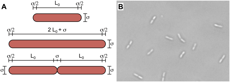
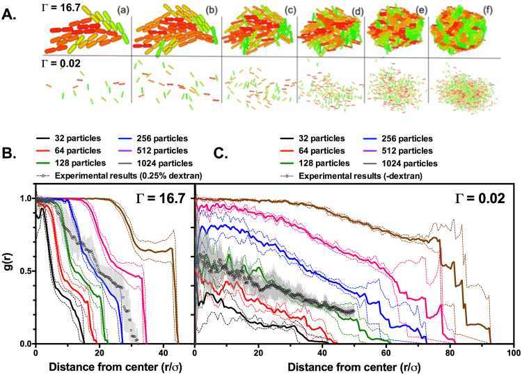
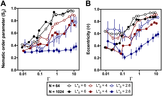
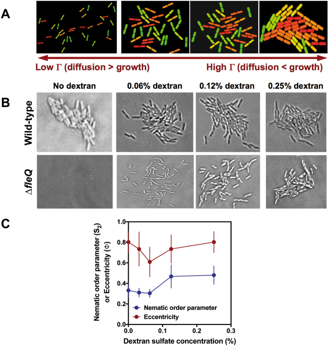
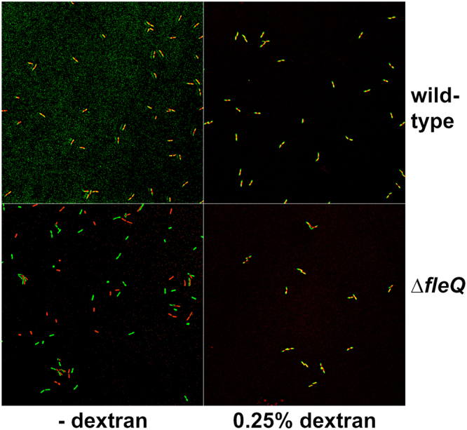
Most bacteria form organized sessile communities, known as biofilms. Their ubiquity and relevance have stimulated the development of efficient mathematical models able to predict biofilm evolution and characteristics at different conditions. Here we present a study of the early stages of bacterial biofilm formation modeled by means of individual cell-based computer simulation. Simulation showed that clusters with different degrees of internal and orientational order were formed as a function of the aspect ratio of the individual particles and the relation between the diffusion and growth rates. Analysis of microscope images of early biofilm formation by the Gram-negative bacterium Pseudomonas putida at varying diffusion rates revealed a good qualitative agreement with the simulation results. Our model is a good predictor of microcolony morphology during early biofilm development, showing that the competition between diffusion and growth rates is a key aspect in the formation of stable biofilm microcolonies.


Conflict of interest statement

The authors declare no competing interests.

Figures

Figure 1
Figure 1
Bacterial cells as spherocylindrical particles. (A) Representation of a particle with its initial elongation L0, an elongated particle prior to division (L = 2 L0 + σ) and two particles immediately after the division (L = L0). The main geometric parameters of our model are shown. (B) Micrograph of P. putida single and dividing cells.
Figure 2
Figure 2
Surface coverage of simulated particle clusters and biofilm microcolonies. (A) Snapshots of 32, 64, 128, 256, 512 or 1024 (a–f) particle clusters with L*0 = 2.6, and Г = 16.7 (top) or 0.02 (bottom). B. and C. Surface coverage profiles g(r) of clusters with L*0 = 2.6, and Г = 16.7 (B) or 0.02 (C). Clusters containing 32, 64, 128, 256, 512 or 1024 particles are denoted by black, red, green, blue, purple and grey lines, respectively. Circles denote surface coverage profiles obtained from MRB52 microcolonies in the presence (B) or in the absence (C) of 0.25% dextran sulfate. Lines and data points represent the average of 10 independent simulations or 10 independent microcolonies containing 117 ± 11 and 65 ± 10 (for the experimental results displayed in B and C, respectively). Dashed lines (simulation data) and error bars (experimental data) represent the standard deviation of the mean.
Figure 3
Figure 3
Typical results from computer simulations. Snapshots showing the shape and internal structure of clusters containing 64 (A) or 1024 (B) particles with aspect ratios L*0 = 2.6, 4 or 6 and variable Г values. Particle color indicates orientation in a scale ranging from green (vertical) to red (horizontal).
Figure 4
Figure 4
Measurement of the nematic order parameter (S2) and eccentricity (Φ) in simulations. (A) Dependence of the nematic order parameter S2 with Г. (B) dependence of the eccentricity Ф with Г. Both plots display simulation results for clusters containing 1024 particles (solid symbols) or 64 particles (open symbols), containing particles with L* = 6 (black), 4 (red) or 2.6. Error bars represent standard deviation of the mean, and are only shown when larger than the corresponding symbol.
Figure 5
Figure 5
Comparison of simulation and live bacterial microcolony microscopy results. (A) Typical snapshots of simulated clusters containing 64 particles of L*0 = 2.6 at different Г values. (B) Micrographs of microcolonies containing ~70 cells of the wild-type and ∆fleQ P. putida strains KT2442 and MRB52 at different dextran sulfate concentrations. (C) Plot representing the experimentally derived eccentricity (Ф) and nematic order parameter (S2) values for colonies of MRB52 grown at different dextran sulfate concentrations. Symbols represent the averages obtained from ten colonies containing 65 ± 9 cells (no dextran sulfate), 74 ± 11 cells (0.031% dextran sulfate), 112 ± 17 cells (0.062% dextran sulfate), 91 ± 19 cells (0.125% dextran sulfate), and 117 ± 8 cells (0.25% dextran sulfate), respectively. Error bars represent standard deviation of the mean.
Figure 6
Figure 6
Effect of dextran sulfate on brownian diffusion of individual wild-type and ∆fleQ P. putida cells. Images are overlays of artificially colored surface-associated wild-type (A) and ∆fleQ (B) cells showing their initial (time = 0; green) and final positions (time = 10 minutes; red). Overlapped positions of motionless cells are indicated in green. Note that overlap may not be perfect due to cell growth during the 10-minute incubation. The complete time-lapse sequences are available as Supplementary videos 1–4.

References

    1. Costerton JW, Lewandowski Z, Caldwell DE, Korber DR, Lappin-Scott HM. Microbial biofilms. Annu. Rev. Microbiol. 1995;49:711–745. doi: 10.1146/annurev.mi.49.100195.003431. - DOI - PubMed
    1. Costerton JW, Stewart PS, Greenberg EP. Bacterial biofilms: a common cause of persistent infections. Science. 1999;284:1318–1322. doi: 10.1126/science.284.5418.1318. - DOI - PubMed
    1. O’Toole GA, Kaplan HB, Kolter R. Biofilm formation as microbial development. Annu. Rev. Microbiol. 2000;54:49–79. doi: 10.1146/annurev.micro.54.1.49. - DOI - PubMed
    1. Monds RD, O’Toole GA. The developmental model of microbial biofilms: ten years of a paradigm up for review. Trends Microbiol. 2009;17:73–87. doi: 10.1016/j.tim.2008.11.001. - DOI - PubMed
    1. Peyton BM, Characklis WG. Microbial biofilms and biofilm reactors. Bioprocess Technol. 1995;20:187–231. - PubMed

Publication types