. 2018 Mar 28;8(1):5294.
doi: 10.1038/s41598-018-23673-z.

Dexras1 is a homeostatic regulator of exercise-dependent proliferation and cell survival in the hippocampal neurogenic niche

Affiliations

Dexras1 is a homeostatic regulator of exercise-dependent proliferation and cell survival in the hippocampal neurogenic niche

Pascale Bouchard-Cannon et al. Sci Rep. .

Abstract

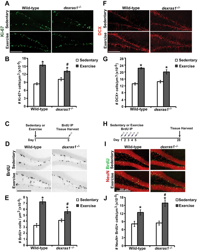
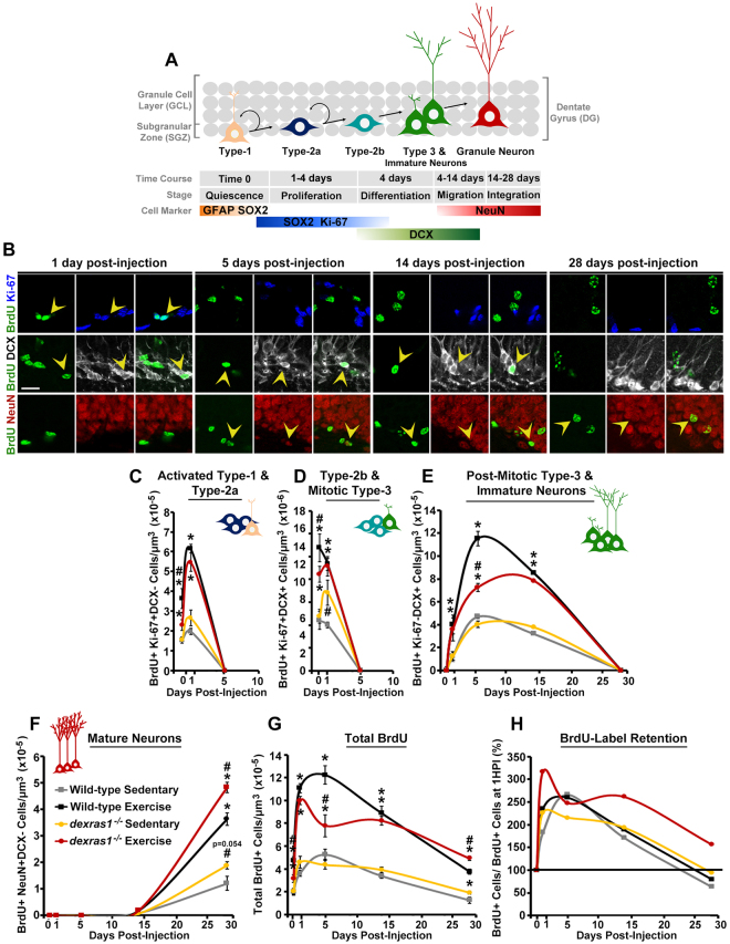
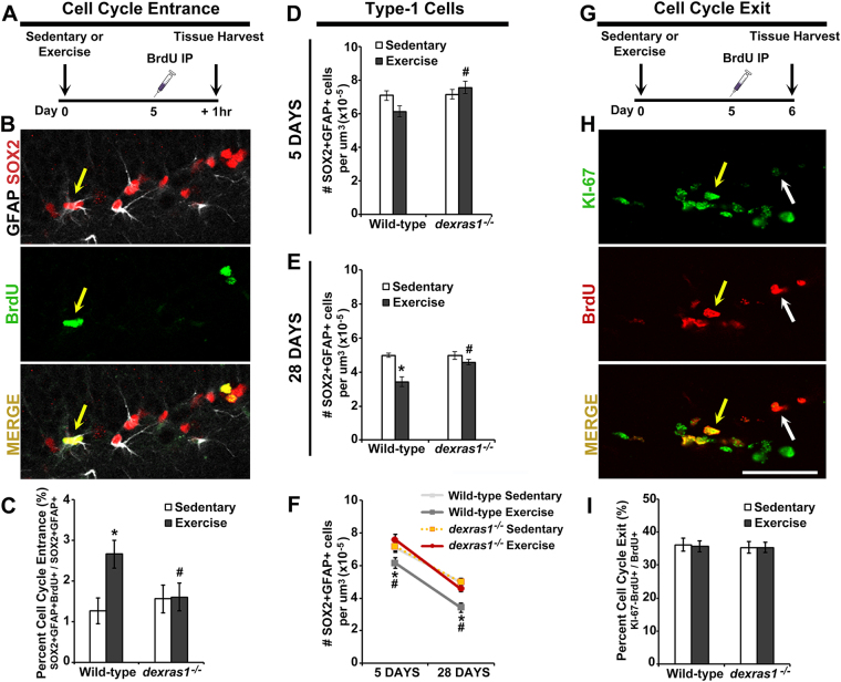
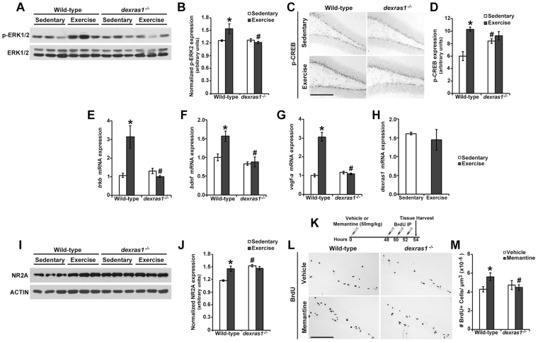
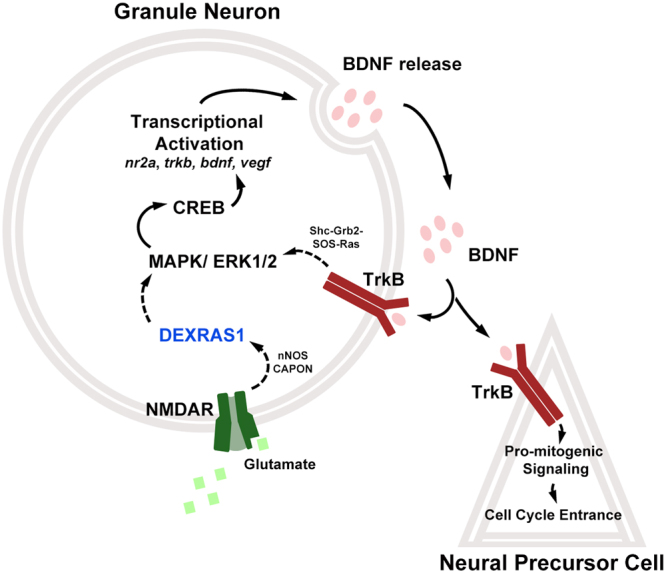
Adult hippocampal neurogenesis is highly responsive to exercise, which promotes the proliferation of neural progenitor cells and the integration of newborn granule neurons in the dentate gyrus. Here we show that genetic ablation of the small GTPase, Dexras1, suppresses exercise-induced proliferation of neural progenitors, alters survival of mitotic and post-mitotic cells in a stage-specific manner, and increases the number of mature newborn granule neurons. Dexras1 is required for exercise-triggered recruitment of quiescent neural progenitors into the cell cycle. Pharmacological inhibition of NMDA receptors enhances SGZ cell proliferation in wild-type but not dexras1-deficient mice, suggesting that NMDA receptor-mediated signaling is dependent on Dexras1. At the molecular level, the absence of Dexras1 abolishes exercise-dependent activation of ERK/MAPK and CREB, and inhibits the upregulation of NMDA receptor subunit NR2A, bdnf, trkB and vegf-a expression in the dentate gyrus. Our study reveals Dexras1 as an important stage-specific regulator of exercise-induced neurogenesis in the adult hippocampus by enhancing pro-mitogenic signaling to neural progenitor cells and modulating cell survival.


Conflict of interest statement

The authors declare no competing interests.

Figures

Figure 1
Figure 1
Dexras1 regulates exercise-induced proliferation of SGZ progenitor cells and survival of newborn neurons. (A) Representative photomicrographs of Ki-67+ cells (green) in the SGZ of wild-type and dexras1−/− mice after 5 days of sedentary or exercise condition. Images are represented as a collapsed z-stack project (30-μm) acquired at 40× magnification. (B) Quantification of the number of Ki-67+ cells per μm3 of SGZ (x10−5). (C) Single BrdU-injection paradigm. Wild-type and dexras1−/− mice received a single BrdU injection after 5 days of sedentary or exercise condition, and tissues were harvested 1 hr post-injection. (D) Representative photomicrographs of BrdU+ cells (black) at day 5 of sedentary or exercise condition acquired at 10× magnification. (E) Quantification of the number of BrdU+ cells per μm3 of SGZ (×10−5). (F) Representative photomicrographs of DCX+ cells (red) from wild-type and dexras1−/− mice following 14 days of sedentary or exercise conditions. Images are represented as a single z-stack image (5-μm) acquired at 40× magnification. (G) Density of DCX+ cells in and adjacent to the SGZ. An area encompassing 2× the thickness of the SGZ was drawn and immunoreactive cells within this region were counted. (H) Label-retaining assay paradigm. Wild-type and dexras1−/− mice received single daily injections of BrdU on days 1 to 5 of sedentary or exercise condition. Tissues were harvested 28 days after the first BrdU injection. (I) Representative photomicrographs of BrdU+ cells (green) and NeuN+ cells (red) in the DG. Images are represented as a collapsed z-stack project (30-μm) acquired at 40× magnification. (J) Quantification of the number of BrdU+NeuN+ cells per μm3 of DG (x10−5). Scale bar = 200 μm. All values represent mean ± standard error. *p < 0.05 vs. sedentary control. #p < 0.05 vs. wild-type control. n = 5–7 per group.
Figure 2
Figure 2
Dexras1 ablation alters the proliferative and survival properties of progenitor cells in a stage-specific manner. (A) Graphical representation of hippocampal neurogenesis and respective markers used to identify the various stage-specific cell types. (B–H) Wild-type and dexras1−/− mice were injected with a single dose of BrdU on day 5 of sedentary or exercise condition. Tissues were harvested after 1 hr (0 DPI) or after 1, 5, 14 or 28 days-post injection (DPI). (B) Representative photomicrographs showing the BrdU label (green) in cells positive for Ki-67 (blue), DCX (white) or NeuN (red) at 1, 5, 14 or 28 DPI. Yellow arrowheads indicate cells with co-localized expression. Images are represented as a single z-stack image (5-μm) acquired at 40× magnification. Scale bar = 20 μm. (C–G) Quantification of the density of BrdU-labeled (C) Ki-67+DCX activated type-1 and type-2a cells, (D) Ki-67+DCX+ type-2b and mitotic type-3 cells, (E) Ki-67DCX+ post-mitotic type-3 cells and immature neurons, and (F) NeuN+DCX- mature neurons. (G) Quantification of the density of all BrdU-labeled cells. X-axis indicates the number of DPI. Data are represented as mean number of cells per μm3 of tissue (x10−6 or x10−5) ± standard error. (H) Percent of BrdU-label retention. Percentage values are calculated as the number BrdU+ cells at each DPI divided by the number of BrdU+ cells at 1 hr post-injection (1 HPI). Wild-type sedentary (grey); wild-type exercise (black); dexras1−/−sedentary (yellow); dexras1−/−exercise (red). *p < 0.05 vs. sedentary control. #p < 0.05 vs. wild-type control. n = 4–6 per group. Refer to Table S1 for cell counts.
Figure 3
Figure 3
Dexras1 is required for exercise-mediated recruitment of type-1 cells into the cell cycle. (A) BrdU-injection paradigm for cell cycle entrance. Wild-type and dexras1−/− mice were placed under sedentary or exercise condition, injected once with BrdU on day 5, and killed for tissue after 1 hr post-injection. (B) Representative photomicrographs of BrdU-labeled (green) cells that co-express GFAP (white) and SOX2 (red). (C) Percent of type-1 cells that recently entered the cell cycle. Values are calculated by dividing the number of BrdU+GFAP+SOX2+ cells by the total number of GFAP+SOX2+ cells in the SGZ. (D–F) Quantification of the number of SOX2+GFAP+ cells per μm3 of SGZ (x10−5) after (D) 5 days or (E) 28 days of sedentary or exercise condition. (F) shows the combined data of both time points. (G) BrdU-injection paradigm for cell cycle exit. Wild-type and dexras1−/− mice were injected once with BrdU on day 5 of sedentary or exercise conditions, and killed for tissue after 24 hr post-injection. (H) Representative photomicrographs of BrdU-labeled (red) cells that co-express Ki-67 (green). (I) Percent of proliferating cells that have exited the cell cycle after 24 hr. Values are calculated by dividing the number of BrdU+Ki-67 cells by the total number of BrdU+ cells in the SGZ. All values represent mean ± standard error. *p < 0.05 vs. sedentary control. #p < 0.05 vs. wild-type control. n = 5–6 per group. Yellow arrows indicate cells with co-localized expression; white arrow shows a BrdU+Ki-67 cell. Images are represented as a single z-stack image (5-μm) acquired at 40× magnification. Scale bar = 50 μm.
Figure 4
Figure 4
Neither dexras1 ablation nor voluntary exercise alters the average S-phase length and cell cycle length of proliferating neural progenitor cells. Wild-type and dexras1−/− mice received a single IdU injection on day 5 of sedentary or exercise condition. For S-phase length calculations, mice received a CldU injection 4 hr post-IdU injection. For cell cycle length calculations, mice received a CldU injection 18 hr post-IdU injection. Hippocampal sections were harvested 45 min post-CldU injection. (A,B) Representative photomicrographs of single- and double-labeled CldU+ (green) and IdU+ (red) cells used for the quantification of (A) S-phase length and (B) cell cycle length. Images are represented as a single z-stack image (5-μm) acquired at 40× magnification. Scale bar = 30 μm. (C–E) Mathematical formulas used for the calculation of (C) S-phase length (Ts), (D) cell cycle length (Tc), and (E) the estimated G1/G2/M-phase combined length. (F) Pie chart representation of the average cell cycle length, S-phase length and estimated G1/G2/M combined length. Values represent mean ± standard error or estimated value. n = 6–9 per group.
Figure 5
Figure 5
Exercise-induced activation of pro-mitogenic signaling cascades is dependent on Dexras1 expression. (A) Representative western blots showing the expression of phospho-ERK1/2 (p-ERK1/2) and total ERK1/2, in the DG of wild-type and dexras1−/−mice after 5 days of sedentary or exercise condition. p-ERK1/2 western blots were optimized for quantification of p42 (ERK2). p44 (ERK1) could be visualized with longer development times (not shown). (B) Quantification of the relative protein abundance of p-ERK2, normalized to total ERK1/2. n = 3 per genotype and condition. (C) Representative photomicrographs of phospho-CREB (p-CREB) immunoreactivity in the SGZ of wild-type and dexras1−/−mice after 5 days of sedentary or exercise condition. Scale bar = 200 μm. (D) Quantification of p-CREB immunoreactive intensity in the SGZ. n = 4–6 per group. (E–H) qRT-PCR analysis of the relative mRNA abundance of (E) trkB, (F) bdnf, (G) vegf-a and (H) dexras1 in the DG of wild-type and dexras1−/−mice after 5 days of sedentary and exercise condition. Values were normalized to gapdh expression. n = 3–4 per genotype and condition. (I) Representative western blot showing the expression of NMDA Receptor 2A (NR2A) and ACTIN in the DG of wild-type and dexras1−/− mice after 5 days of sedentary or exercise condition. (J) Quantification of the relative protein abundance of NR2A normalized to actin. n = 3 per genotype and condition. (K) Memantine and BrdU-injection paradigm. Sedentary wild-type and dexras1−/−mice were injected once with memantine or vehicle prior to receiving 3 injections of BrdU spaced 2 hr apart starting 48 hr after memantine administration. Tissues were harvested 2 hr after the last BrdU injection. (L) Representative photomicrographs of BrdU+ (black) cells in the SGZ of memantine- or vehicle-treated mice. Scale bar = 200 μm. (M) Quantification of the number of BrdU+ cells per μm3 of SGZ (x10−5) in memantine- or vehicle-treated mice. n = 4–5 per group. All values represent mean ± standard error. *p < 0.05 vs. sedentary control. #p < 0.05 vs. wild-type control.
Figure 6
Figure 6
Hypothetical model for Dexras1-mediated recruitment of quiescent neural progenitor cells into the cell cycle. Hypothetical model depicting the role of Dexras1 in the regulation of exercise-induced neurogenesis. The model asserts that Dexras1 couples NMDA receptor activation to MAPK/ERK signaling in granule neurons, leading to CREB-mediated transcriptional activation of pro-mitogenic genes including bdnf. BDNF is released and acts on type-1 cells to promote cell cycle entrance. It can also act on granule neurons to further bolster the activity of the NMDAR-MAPK/ERK-CREB cascade.

Similar articles

Cited by

References

    1. Stolp HB, Molnár Z. Neurogenic niches in the brain: help and hindrance of the barrier systems. Front. Neurosci. 2015;9:1–7. doi: 10.3389/fnins.2015.00020. - DOI - PMC - PubMed
    1. Seri B, Garcia-Verdugo JM, Mcewen BS, Alvarez-buylla A. Astrocytes Give Rise to New Neurons in the Adult Mammalian Hippocampus. J. Neurosci. 2001;21:7153–7160. - PMC - PubMed
    1. Babu H, Cheung G, Kettenmann H, Palmer TD, Kempermann G. Enriched Monolayer Precursor Cell Cultures from Micro- Dissected Adult Mouse Dentate Gyrus Yield Functional Granule Cell-Like Neurons. PLoS One. 2007;2:1–12. doi: 10.1371/journal.pone.0000388. - DOI - PMC - PubMed
    1. Kempermann G, Song H, Gage FH. Neurogenesis in the AdultHippocampus. Cold Spring Harb. Perspect. Biol. 2015;7:1–14. - PMC - PubMed
    1. Steiner B, et al. Type-2 Cells as Link Between Glial and Neuronal Lineage in Adult Hippocampal Neurogenesis. Glia. 2006;54:805–814. doi: 10.1002/glia.20407. - DOI - PubMed

Publication types

MeSH terms

Substances

Grants and funding