. 2018 Mar 20:11:95-109.
doi: 10.2147/JIR.S152835. eCollection 2018.

Glucagon-like peptide-1 exerts anti-inflammatory effects on mouse colon smooth muscle cells through the cyclic adenosine monophosphate/nuclear factor-κB pathway in vitro

Affiliations

Glucagon-like peptide-1 exerts anti-inflammatory effects on mouse colon smooth muscle cells through the cyclic adenosine monophosphate/nuclear factor-κB pathway in vitro

Ahmed Al-Dwairi et al. J Inflamm Res. .

Erratum in

Abstract

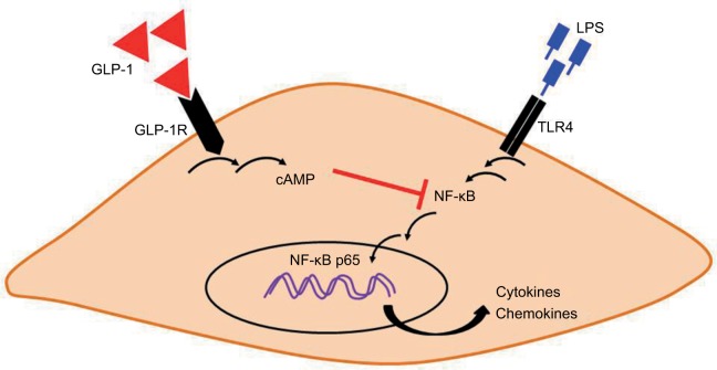
Background: Intestinal smooth muscle cells (SMCs) undergo substantial morphological, phenotypic, and contractile changes during inflammatory bowel disease (IBD). SMCs act as a source and target for different inflammatory mediators, however their role in IBD pathogenesis is usually overlooked. Glucagon-like peptide-1 (GLP-1) is an incretin hormone reported to exert multiple anti-inflammatory effects in different tissues including the gastrointestinal tract through various mechanisms.

Aim: The aim of this research is to explore the effect of GLP-1 analog exendin-4 on the expression and secretion of inflammatory markers from mouse colon smooth muscle cells (CSMCs) after stimulation with lipopolysaccharide (LPS).

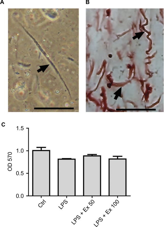
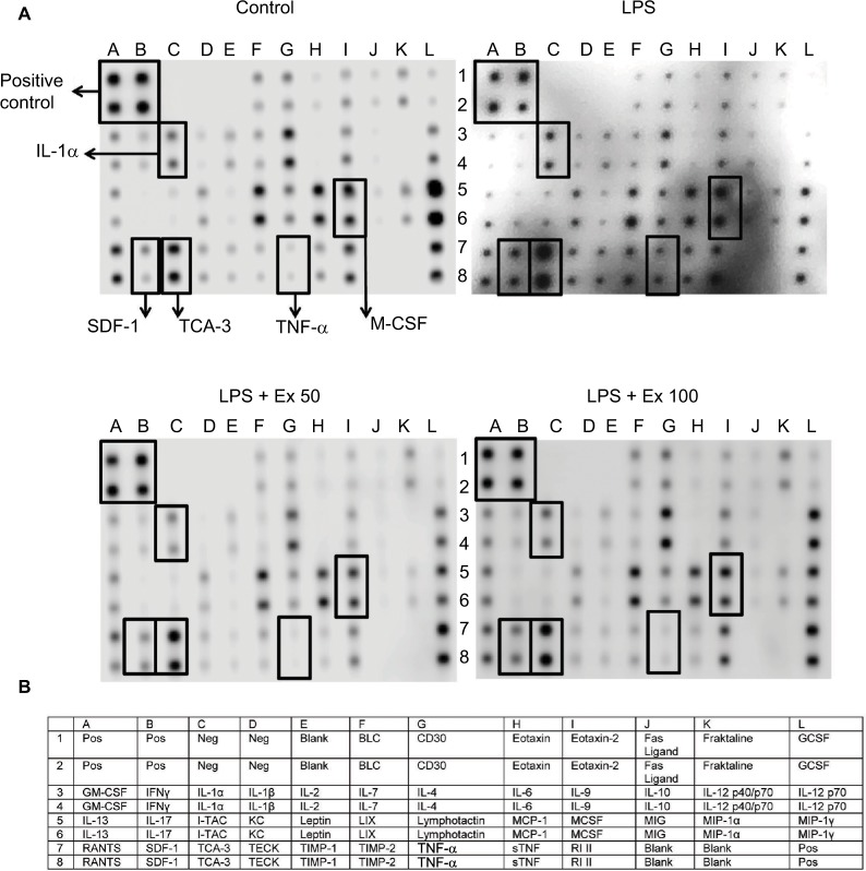
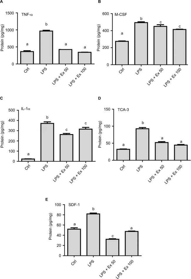
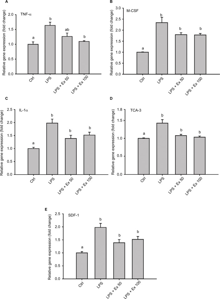
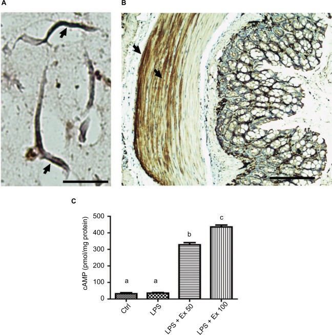
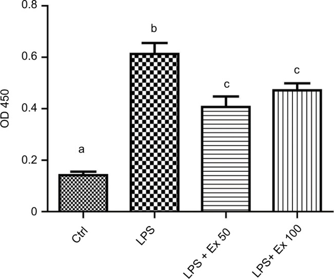
Materials and methods: Freshly isolated CSMCs from male BALB/c mice were cultured in DMEM and treated with vehicle, LPS (1 μg/mL), LPS+exendin-4 (50 nM), or LPS+exendin-4 (100 nM) for 24 h. Expression of inflammatory cytokines was then evaluated by antibody array membrane.

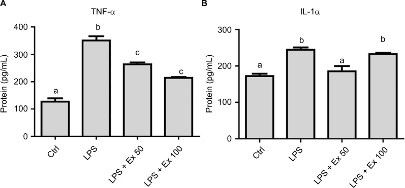
Results: CSMCs showed basal expression of several cytokines which was enhanced with the induction of inflammation by LPS. However, exendin-4 (50 and 100 nM) significantly (p<0.05) reduced the expression of multiple cytokines including tumor necrosis factor-α (TNF-α), interleukin-1α (IL-1α), T cell activation gene-3 (TCA-3), stromal cell-derived factor-1 (SDF-1), and macrophage colony stimulating factor (M-CSF). To confirm these results, expression of these cytokines was further assessed by enzyme-linked immunosorbent assay and real-time polymerase chain reaction and similar results were also observed. Moreover, secretion of TNF-α and IL1-α into the conditioned media was significantly downregulated by exendin-4 when compared to LPS-treated cells. Furthermore, LPS increased NF-κB phosphorylation, while exendin-4 significantly reduced levels of NF-κB phosphorylation.

Conclusion: These data indicate that GLP-1 analogs can exert significant anti-inflammatory effects on CSMCs and can potentially be used as an adjunct treatment for inflammatory bowel conditions.

Keywords: GLP-1; inflammation; inflammatory bowel disease; smooth muscle.


Conflict of interest statement

Disclosure The authors report no conflicts of interest in this work.

Figures

Figure 1
Figure 1
Identification of mouse CSMCs. (A) Single CSMC appeared in spindle shape (arrow) under phase contrast microscopy. Cells were viewed via inverted Nikon TMS-F microscope, and the image captured with a Canon digital camera. (B) Immunohistochemical staining of paraffin-embedded mouse CSMCs (arrows) using anti-h1-calponin antibody. (C) Cell viability assay for cells treated with vehicle (Ctrl), LPS, LPS+exendin-4 (50 nM), or LPS+exendin-4 (100 nM). Pictures were viewed at 20× magnification, scale bar=50 μm. Abbreviations: CSMC, colon smooth muscle cell; Ex 50, exendin-4 (50 nM); Ex 100, exendin-4 (100 nM); LPS, lipopolysaccharide; OD, optical density; Ctrl, control.
Figure 2
Figure 2
Effect of exendin-4 treatment on the expression of inflammatory cytokines by mouse CSMCs using antibody array membrane. (A) Freshly isolated CSMCs from 20 male Balb/C mice were cultured in DMEM and divided into four treatment groups: control, LPS, LPS+exendin-4 (50 nM), and LPS+exendin-4 (100 nM). After 24 h of incubation the cells were collected, lysed, and incubated with the antibody array membrane. There was a basal expression of various cytokines by the normal CSMCs which was increased once inflammation was induced by LPS. Exendin-4 at both concentrations inhibited the overproduction of cytokines induced by inflammation. A visual comparison between the four arrays performed by two independent investigators and by ImageJ software (National Institutes of Health) revealed that five cytokines were especially affected by the treatment: tumor necrosis factor-α (TNF-α), interleukin-1α (IL-1α), T cell activation gene-3 (TCA-3), stromal cell-derived factor-1 (SDF-1), and macrophage colony stimulating factor (M-CSF). (B) List of cytokines and chemokines represented by the antibody array membrane (Cat. No. ab133999; Abcam Inc.). Abbreviations: CSMC, colon smooth muscle cell; Ex 50, exendin-4 (50 nM); Ex 100, exendin-4 (100 nM); GCSF, granulocyte colony stimulating factor; GM-CSF, granulocyte– macrophage colony stimulating factor; IFN, interferon; IL, interleukin; LPS, lipopolysaccharide; Pos, positive; Neg, negative.
Figure 3
Figure 3
Effect of exendin-4 treatment on the expression of inflammatory cytokines by mouse CSMCs using ELISA. Freshly isolated CSMCs from 20 male Balb/C mice were cultured in DMEM in 6-well plates and divided into four treatment groups (n=3/group): control (Ctrl), LPS, LPS+exendin-4 (50 nM), and LPS+exendin-4 (100 nM). After 24 h of incubation the cells were collected and lysed. Levels of different cytokines in the lysate were assayed using specific ELISA kits. Bar graphs represent levels of: (A) tumor necrosis factor-α (TNF-α), (B) macrophage colony stimulating factor (M-CSF), (C) interleukin-1α (IL-1α), (D) T cell activation gene-3 (TCA-3), and (E) stromal cell-derived factor-1 (SDF-1). Different lowercase letters indicate significant statistical difference between groups where p<0.05. Values shown are representative of three independent experiments performed in triplicates. Bars represent mean±standard error of the mean. Abbreviations: CSMC, colon smooth muscle cell; ELISA, enzyme-linked immunosorbent assay; Ex 50, exendin-4 (50 nM); Ex 100, exendin-4 (100 nM); LPS, lipopolysaccharide.
Figure 4
Figure 4
Effect of exendin-4 treatment on the secretion of inflammatory cytokines by mouse CSMCs into the conditioned media using ELISA. Freshly isolated CSMCs from 20 male Balb/C mice were cultured in DMEM in 6-well plates and divided into four treatment groups (n=3/group): control (Ctrl), LPS, LPS+exendin-4 (50 nM), and LPS+exendin-4 (100 nM). After 24 h of incubation the cells were collected and lysed as mentioned previously, and the conditioned media were stored for later analysis at −20 °C. Levels of different cytokines in the conditioned media were assayed using specific ELISA kits. Bar graphs represent levels of: (A) tumor necrosis factor-α (TNF-α) and (B) interleukin-1α (IL-1α). Different lowercase letters indicate significant statistical difference between groups where p<0.05. Values shown are representative of three independent experiments performed in triplicates. Bars represent mean±standard error of the mean. Abbreviations: CSMC, colon smooth muscle cell; ELISA, enzyme-linked immunosorbent assay; Ex 50, exendin-4 (50 nM); Ex 100, exendin-4 (100 nM); LPS, lipopolysaccharide.
Figure 5
Figure 5
Effect of exendin-4 treatment on mRNA abundance of inflammatory cytokines in mouse CSMCs using RT-PCR. Freshly isolated CSMCs from 20 male Balb/C mice were cultured in DMEM in 6-well plates and divided into four treatment groups (n=3/group): control (Ctrl), LPS, LPS+exendin-4 (50 nM), and LPS+exendin-4 (100 nM). After 12 h of incubation the cells were collected and RNA was extracted as mentioned previously. Relative gene expression of different cytokines in the treated cell was assayed using RT-PCR. Bar graphs represent gene expression levels of: (A) tumor necrosis factor-α (TNF-α), (B) macrophage colony stimulating factor (M-CSF), (C) interleukin-1α (IL-1α), (D) T cell activation gene-3 (TCA-3), and (E) stromal cell-derived factor-1 (SDF-1). Different lowercase letters indicate significant statistical difference between groups where p<0.05. Values shown are representative of two independent experiments performed in triplicates. Bars represent mean±standard error of the mean. Abbreviations: CSMC, colon smooth muscle cell; Ex 50, exendin-4 (50 nM); Ex 100, exendin-4 (100 nM); LPS, lipopolysaccharide; RT-PCR, real-time/reverse transcription polymerase chain reaction.
Figure 6
Figure 6
Expression of GLP-1 receptor on mouse CSMCs. (A) Micrograph depicting immunohistochemical staining of GLP-1R in mouse isolated single CSMCs, scale bar=40 μM. Arrows show cytoplasmic and membranous staining indicative of GLP-1R expression. (B) Micrograph depicting immunohistochemical staining of GLP-1R in mouse mid-colon section, scale bar=100 μM. Arrows show GLP-1R staining in colon muscularis layer, indicating expression of GLP-1R in CSMCs. (C) cAMP levels in CSMCs treated with: control (Ctrl), LPS, LPS+exendin-4 (50 nM), and LPS+exendin-4 (100 nM) for 10 min. Different lowercase letters indicate significant statistical difference between groups where p<0.05. Abbreviations: cAMP, cyclic adenosine monophosphate; CSMC, colon smooth muscle cell; Ex 50, exendin-4 (50 nM); Ex 100, exendin-4 (100 nM); LPS, lipopolysaccharide.
Figure 7
Figure 7
Exendin-4 reduces LPS-induced nuclear NF-κB phosphorylation. Freshly isolated CSMCs from 20 male Balb/C mice were cultured in DMEM in 6-well plates and divided into four treatment groups (n=3/group): control (Ctrl), LPS, LPS+exendin-4 (50 nM), and LPS+exendin-4 (100 nM). After 24 h of incubation the cells were collected and lysed. Total nuclear proteins were extracted using specific nuclear extraction kit. Nuclear NF-κB p65 pS536 levels were assayed in the lysate using a specific ELISA kit. Different lowercase letters indicate significant statistical difference between groups where p<0.05. Values shown are representative of two independent experiments performed in triplicates. Bars represent mean±standard error of the mean. Abbreviations: CSMC, colon smooth muscle cell; Ex 50, exendin-4 (50 nM); Ex 100, exendin-4 (100 nM); LPS, lipopolysaccharide; NF, nuclear factor; OD, optical density.
Figure 8
Figure 8
A proposed integrative model for how GLP-1 analog exendin-4 may suppress LPS-induced inflammatory gene expression in mouse CSMCs. Binding of exendin-4 to cell surface GLP-1R may activate adenylyl cyclase and increase the formation of intracellular second messenger cAMP. cAMP can interfere with LPS-induced NF-κB activation, phosphorylation, and translocation to the nucleus, which in turn suppress inflammatory cytokine and chemokine expression. Abbreviations: cAMP, cyclic adenosine monophosphate; CSMC, colon smooth muscle cell; GLP-1, glucagon-like peptide-1; GLP-1R, GLP-1 receptor; LPS, lipopolysaccharide; NF, nuclear factor; TLR, toll-like receptor.

References

    1. Abraham C, Cho JH. Inflammatory bowel disease. N Engl J Med. 2009;361(21):2066–2078. - PMC - PubMed
    1. Click B, Binion DG, Anderson AM. Predicting costs of care for patients with inflammatory bowel diseases. Clin Gastroenterol Hepatol. 2017;15(3):393–395. - PMC - PubMed
    1. Ott C, Schölmerich J. Extraintestinal manifestations and complications in IBD. Nat Rev Gastroenterol Hepatol. 2013;10(10):585–595. - PubMed
    1. Fakhoury M, Negrulj R, Mooranian A, Al-Salami H. Inflammatory bowel disease: clinical aspects and treatments. J Inflamm Res. 2014;7:113–120. - PMC - PubMed
    1. Ananthakrishnan AN, Donaldson T, Lasch K, Yajnik V. Management of inflammatory bowel disease in the elderly patient: challenges and opportunities. Inflamm Bowel Dis. 2017;23(6):882–893. - PMC - PubMed