Case Reports
. 2018 Mar 9:9:83.
doi: 10.3389/fendo.2018.00083. eCollection 2018.

Hypophysitis following Treatment with Ustekinumab: Radiological and Pathological Findings

Affiliations
Case Reports

Hypophysitis following Treatment with Ustekinumab: Radiological and Pathological Findings

Ana M Ramos-Leví et al. Front Endocrinol (Lausanne). .

Abstract

Context: Ustekinumab is a human IgG1 monoclonal antibody that targets interleukin (IL)-12 and IL-23, which may be useful in the treatment of autoimmune conditions such as psoriasis, psoriatic arthritis, and Crohn's disease. Hypophysitis is an immune-derived inflammatory condition of the pituitary gland that may lead to pituitary dysfunction. With the increasing use of immunotherapy, it is possible that this and other new immune-related adverse events (IRAEs) arise, although the mechanisms involved are still incompletely defined.

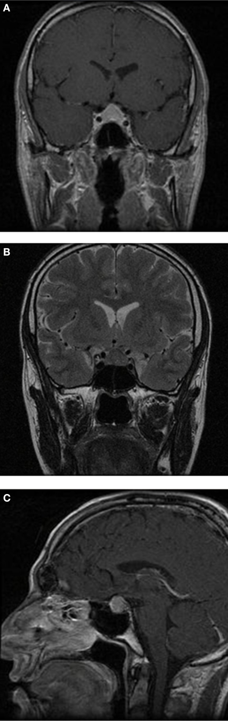
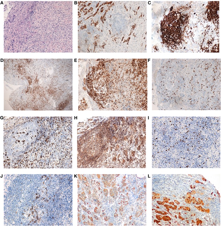
Case description: A 35-year-old male, with a previous history of severe plaque-psoriasis who had started treatment with ustekinumab 4 months before, complained of progressive and persistent headache. Brain magnetic resonance imaging (MRI) was unremarkable. One year later, a new MRI was performed due to headache persistence, which revealed a homogenous and diffuse pituitary enlargement, with suprasellar extension and optic chiasm involvement, blurring of the pituitary stalk, absence of clear differentiation between the anterior and posterior lobes, and no signs of hemorrhage or adenomas. Endocrine evaluation was consistent with panhypopituitarism. Work-up of infiltrative and infectious diseases was negative. Follow-up MRI revealed an increase in the pituitary enlargement and transsphenoidal surgery was performed. Pathological findings revealed an intense fibrosis and a chronic inflammatory infiltrate, but no evidence of adenoma, granuloma, or acid fast bacilli. Immunohistochemical staining showed a combined T-cell (CD3+, CD4+) and B-cell (CD19+, CD20+) phenotype.

Conclusion: We suggest a novel IRAE of ustekinumab, with full radiological and immunopathological iconography, which may be mediated by the complex interaction between different immunological processes.

Keywords: autoimmunity; hypophysitis; pituitary; psoriasis; ustekinumab.


Figures

Figure 1
Figure 1
Brain magnetic resonance imaging (MRI) sections. Brain MRI, showing a homogenous and diffuse pituitary enlargement of up to 1.1 cm, which extended to the suprasellar region and contacted the optic chiasm, blurring of the pituitary stalk, absence of clear differentiation between the anterior and posterior lobes, and no signs of hemorrhage or adenomas. (A) Frontal T1-weighted after gadolinium; (B) frontal T2-weighted; and (C) sagittal T1-weighted after gadolinium.
Figure 2
Figure 2
Pituitary gland serial sections from a representative area of the patient’s pituitary biopsy. Pituitary gland serial sections from a representative area of the patient’s pituitary biopsy, showing the most relevant immune-histopathological findings. Note the germinal center consisting of CD20+ B cells and peripheral CD3+ T cells (classified as T lymphocytes). Both CD4+ (helper) and CD8+ (cytotoxic) lymphocytes were found, as well as abundant macrophages (CD68+). T-regulatory cells were present in germinal centers and in the periphery (Foxp3+ and PD1+). Interleukin (IL)-17 staining was found not only in infiltrating lymphocytes but also in cells of pituitary origin in both hypophysitis and normal pituitary (data not shown). (A) Staining by hematoxylin–eosin; (B) synaptophysin; (C) CD20 (note how CD20+ cells form an aggregate, resembling a lymphoid follicle); (D) CD3; (E) CD4; (F) CD8; (G) CD68; (H) IL-17; (I) FOXp3; (J) PD1; (K) GH; and (L) prolactin. Sections (A–H) are shown as 10× and sections (I–L) as 20×.

References

    1. Hsu L, Snodgrass BT, Armstrong AW. Antidrug antibodies in psoriasis: a systematic review. Br J Dermatol (2014) 170:261–73.10.1111/bjd.12654 - DOI - PubMed
    1. Leung Y, Panaccione R. Update on ustekinumab for the treatment of Crohn’s disease. Gastroenterol Clin North Am (2014) 43:619–30.10.1016/j.gtc.2014.05.013 - DOI - PubMed
    1. Faje A. Immunotherapy and hypophysitis: clinical presentation, treatment, and biologic insights. Pituitary (2016) 19:82–92.10.1007/s11102-015-0671-4 - DOI - PubMed
    1. Joshi MN, Whitelaw BC, Palomar MT, Wu Y, Carroll PV. Immune checkpoint inhibitor-related hypophysitis and endocrine dysfunction: clinical review. Clin Endocrinol (Oxf) (2016) 85:331–9.10.1111/cen.13063 - DOI - PubMed
    1. Deshpande V, Zen Y, Chan JK, Yi EE, Sato Y, Yoshino T, et al. Consensus statement on the pathology of IgG4-related disease. Mod Pathol (2012) 25:1181–92.10.1038/modpathol.2012.72 - DOI - PubMed

Publication types