Review
. 2018 Mar 12:9:395.
doi: 10.3389/fmicb.2018.00395. eCollection 2018.

Signals Involved in Regulation of Hepatitis C Virus RNA Genome Translation and Replication

Affiliations
Review

Signals Involved in Regulation of Hepatitis C Virus RNA Genome Translation and Replication

Michael Niepmann et al. Front Microbiol. .

Abstract

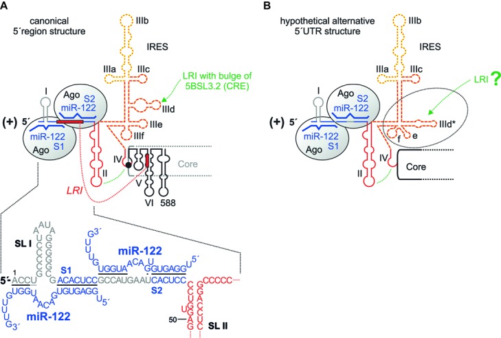
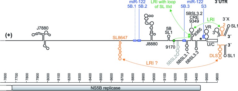
Hepatitis C virus (HCV) preferentially replicates in the human liver and frequently causes chronic infection, often leading to cirrhosis and liver cancer. HCV is an enveloped virus classified in the genus Hepacivirus in the family Flaviviridae and has a single-stranded RNA genome of positive orientation. The HCV RNA genome is translated and replicated in the cytoplasm. Translation is controlled by the Internal Ribosome Entry Site (IRES) in the 5' untranslated region (5' UTR), while also downstream elements like the cis-replication element (CRE) in the coding region and the 3' UTR are involved in translation regulation. The cis-elements controlling replication of the viral RNA genome are located mainly in the 5'- and 3'-UTRs at the genome ends but also in the protein coding region, and in part these signals overlap with the signals controlling RNA translation. Many long-range RNA-RNA interactions (LRIs) are predicted between different regions of the HCV RNA genome, and several such LRIs are actually involved in HCV translation and replication regulation. A number of RNA cis-elements recruit cellular RNA-binding proteins that are involved in the regulation of HCV translation and replication. In addition, the liver-specific microRNA-122 (miR-122) binds to two target sites at the 5' end of the viral RNA genome as well as to at least three additional target sites in the coding region and the 3' UTR. It is involved in the regulation of HCV RNA stability, translation and replication, thereby largely contributing to the hepatotropism of HCV. However, we are still far from completely understanding all interactions that regulate HCV RNA genome translation, stability, replication and encapsidation. In particular, many conclusions on the function of cis-elements in HCV replication have been obtained using full-length HCV genomes or near-full-length replicon systems. These include both genome ends, making it difficult to decide if a cis-element in question acts on HCV replication when physically present in the plus strand genome or in the minus strand antigenome. Therefore, it may be required to use reduced systems that selectively focus on the analysis of HCV minus strand initiation and/or plus strand initiation.

Keywords: HCV; cis-element; microRNA-122; replication; untranslated region.


Figures

FIGURE 1
FIGURE 1
Hepatitis C virus RNA genome replication. The hepatitis C virus (HCV) (+) strand RNA genome (top) is shown with the highly structured 5′ and 3′ untranslated regions (UTRs) and additional RNA secondary structures as mentioned in the main text. The polyprotein start codon AUG is shown as filled circle in stem-loop (SL) IV of the IRES, the polyprotein stop codon as asterisk. The polyprotein is co- and post-translationally processed into the mature structural and non-structural (NS) proteins. The NS3-NS5B proteins form the replication complex with the NS5B protein as the viral RNA dependent RNA polymerase (RdRP, replicase). Stimulatory (green) and inhibitory (red) long-range RNA-RNA interactions (LRIs) and binding sites for the liver-specific microRNA-122 (miR-122) (blue boxes) are shown. The HCV genome replicates by producing a minus strand (-) antigenome (bottom), which in turn is used as the template for progeny plus strand production. RNA secondary structures are named as in the main text. CRE, cis-replication element, DLS, dimerization linkage sequence; IRES, internal ribosome entry site; U/C, poly(U/C) tract; VR, variable region. The drawings of the RNA secondary structures are not to scale in relation to genome length.
FIGURE 2
FIGURE 2
The HCV 5′ region. (A) The canonical HCV 5′ UTR structure with the miR-122 binding sites S1 and S2, bound by Argonaute (Ago) protein. The region of the IRES that binds the small ribosomal 40S subunit in the SL III domain with the important SL IIId is shown by a red broken line, the eIF3 binding site with the important SL IIIb with a yellow dotted line. The SL II interacts with the SL IV to place the AUG into the mRNA entry channel of the 40S ribosomal subunit. SLs V, VI and 588 are located in the core protein coding region. The seed region of miR-122 (nucleotides 2–7 or 2–8) binds to the target sequence (A)CACUCC, and the miR-122 supplementary region binds to a variable number of target nucleotides. (B) A hypothetical alternative structure of the IRES in which the SL IIId region is largely rearranged (Fricke et al., 2015). This predicted structure is conserved among HCV isolates and is as thermodynamically stable as the canonical structure.
FIGURE 3
FIGURE 3
The HCV genome 3′ region. The NS5B sequence is shown with nucleotide numbers. The NS5B stop codon is shown by an asterisk, and the 3′ X region is shown in its two experimentally validated alternative structures. The miR-122 binding sites are shown as blue boxes, with the first non-conserved site 5B.1 with a dotted box and the other conserved two sites in the NS5B region (5B.2 and 5B.3) and the miR-122 target site in the 3′ UTR (S3) with solid boxes (Fricke et al., 2015). Elements suspected to be involved in RNA genome packaging are shown in orange.
FIGURE 4
FIGURE 4
Long-range interactions of the HCV genome ends mediated by trans-acting factors. The small ribosomal 40S subunit interacts with the lower part of the SL III domain in the HCV 5′ UTR and with the HCV 3′ UTR, preferentially with the poly(U/C) tract (U/C). The NF90/NF45 complex interacts with the upper part of the SL III domain in the 5′ UTR and with the SL 3 in the 3′ UTR. PCBP2 interacts with each two sites in the 5′ UTR and in the 3′ UTR. IGF2BP1 (or IMP1) interacts with the 3′ region of the IRES and with the poly(U/C) tract of the 3′ UTR. A hypothetical interaction (shown by gray dotted lines) may also involve hnRNP L (which binds the 3′ region of the IRES) and PTB [which binds the poly(U/C) tract]. In addition, cooperative aggregation of miR-122/Ago complexes may also be hypothesized to contribute to the RNA end-to-end communication (not illustrated in the figure).
FIGURE 5
FIGURE 5
Elements in the 3′ region of the HCV minus strand antigenome involved in plus strand synthesis. The black and gray lines and structures show the 3′ region of the HCV minus strand antigenome. A contribution to plus strand initiation shown by in vitro-assays is indicated by boxes with solid green lines (SL I′, SL IIIb′), a negative influence indicated by these in vitro-assays is indicated by a box with a red broken line (SL IIz′). Sequences that were characterized by replicon studies to have a positive influence on RNA replication are boxed with dotted green lines (SL IIz′ and SL IIy′). The region which is not boxed is variable in RNA secondary structure among the predictions (see text), and no study showed a functional contribution of this region to replication. A possible relevance of the upstream structures SL VI′ and 588′ as present in the minus strand is not clear.

Similar articles

Cited by

References

    1. Adams R. L., Pirakitikulr N., Pyle A. M. (2017). Functional RNA structures throughout the Hepatitis C Virus genome. Curr. Opin. Virol. 24 79–86. 10.1016/j.coviro.2017.04.007 - DOI - PMC - PubMed
    1. Alvarez D. E., Lodeiro M. F., Luduena S. J., Pietrasanta L. I., Gamarnik A. V. (2005). Long-range RNA-RNA interactions circularize the dengue virus genome. J. Virol. 79 6631–6643. 10.1128/JVI.79.11.6631-6643.2005 - DOI - PMC - PubMed
    1. Angus A. G., Dalrymple D., Boulant S., McGivern D. R., Clayton R. F., Scott M. J., et al. (2010). Requirement of cellular DDX3 for hepatitis C virus replication is unrelated to its interaction with the viral core protein. J. Gen. Virol. 91 122–132. 10.1099/vir.0.015909-0 - DOI - PMC - PubMed
    1. Ariumi Y., Kuroki M., Kushima Y., Osugi K., Hijikata M., Maki M., et al. (2011). Hepatitis C virus hijacks P-body and stress granule components around lipid droplets. J. Virol. 85 6882–6892. 10.1128/JVI.02418-10 - DOI - PMC - PubMed
    1. Astier-Gin T., Bellecave P., Litvak S., Ventura M. (2005). Template requirements and binding of hepatitis C virus NS5B polymerase during in vitro RNA synthesis from the 3′-end of virus minus-strand RNA. FEBS J. 272 3872–3886. 10.1111/j.1742-4658.2005.04804.x - DOI - PubMed