. 2018 Mar 12:9:346.
doi: 10.3389/fimmu.2018.00346. eCollection 2018.

Mucosal Delivery of Fusion Proteins with Bacillus subtilis Spores Enhances Protection against Tuberculosis by Bacillus Calmette-Guérin

Affiliations

Mucosal Delivery of Fusion Proteins with Bacillus subtilis Spores Enhances Protection against Tuberculosis by Bacillus Calmette-Guérin

Alastair Copland et al. Front Immunol. .

Abstract

Tuberculosis (TB) is the most deadly infectious disease in existence, and the only available vaccine, Bacillus Calmette-Guérin (BCG), is almost a century old and poorly protective. The immunological complexity of TB, coupled with rising resistance to antimicrobial therapies, necessitates a pipeline of diverse novel vaccines. Here, we show that Bacillus subtilis spores can be coated with a fusion protein 1 ("FP1") consisting of Mycobacterium tuberculosis (Mtb) antigens Ag85B, ACR, and HBHA. The resultant vaccine, Spore-FP1, was tested in a murine low-dose Mtb aerosol challenge model. Mice were primed with subcutaneous BCG, followed by mucosal booster immunizations with Spore-FP1. We show that Spore-FP1 enhanced pulmonary control of Mtb, as evidenced by reduced bacterial burdens in the lungs. This was associated with elevated antigen-specific IgG and IgA titers in the serum and lung mucosal surface, respectively. Spore-FP1 immunization generated superior antigen-specific memory T-cell proliferation in both CD4+ and CD8+ compartments, alongside bolstered Th1-, Th17-, and Treg-type cytokine production, compared to BCG immunization alone. CD69+CD103+ tissue resident memory T-cells (Trm) were found within the lung parenchyma after mucosal immunization with Spore-FP1, confirming the advantages of mucosal delivery. Our data show that Spore-FP1 is a promising new TB vaccine that can successfully augment protection and immunogenicity in BCG-primed animals.

Keywords: adjuvants; immunity; spores; tuberculosis; vaccine.


Figures

Figure 1
Figure 1
Spore-FP1 protects against aerosol Mycobacterium tuberculosis (Mtb) challenge. Mice received a Bacillus Calmette-Guérin subcutaneous prime (except the PBS control group) followed by two intranasal boosts with Spore-FP1. After 3 weeks, bacterial burdens in the lungs and spleens were quantified by CFU counting on 7H11 plates across three dilution ranges. (A) Mice were immunized with Spore-FP1 alone. (B) Mice were immunized with Spore-FP1 in combination with the adjuvant poly(I:C). Results are expressed as mean ± SEM. Data are derived from n = 4–7 individual mice. Significance was tested against the by one-way ANOVA with Tukey’s posttest, *p < 0.05, **p < 0.01, ***p < 0.001, and ****p < 0.0001.
Figure 2
Figure 2
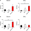
Enhanced humoral immunity caused by Spore-FP1. Immunized mice were tested for the presence of antigen-specific IgG in the serum (1:1,000 dilution) and IgA in the BAL (1 mL PBS flush; 1:10 dilution) by ELISA, with optical density read at 450 nm in duplicate. (A) Levels of IgG and IgA specific to Ag85B. (B) Levels of IgG and IgA specific to ACR. Results are expressed as mean ± SEM. Data shown are derived from n = 3 individual mice and are representative of two independent experiments. Significance was tested against the unstimulated control by one-way ANOVA with Tukey’s posttest, *p < 0.05 and **p < 0.01.
Figure 3
Figure 3
Enhanced T-cell proliferation due to Spore-FP1. Splenocytes were incubated in technical duplicates with 5 µg/mL recall antigen for 5–6 days and proliferation was measured by Ki67 staining. A gating strategy of live cells→single cells→CD3+→CD4+/CD8+ was used, followed by gating for Ki67+ cells and determination of memory cell phenotype by expression of CD44 and CD62L. Results are expressed as mean ± SEM. Data are derived from n = 3 pooled spleens per group.
Figure 4
Figure 4
Cytokine profiles during splenocyte antigen recall. Splenocytes from immunized mice were stimulated in technical duplicates with 5 µg/mL recall antigen for 5–6 days and T-cell cytokines were measured by multiplex flow cytometry. Results are expressed as mean ± SEM. Data are derived from n = 3 pooled spleens per group.
Figure 5
Figure 5
Spore-FP1 induces enrichment of tissue resident memory cells. Mice were first immunized with Bacillus Calmette-Guérin for 6 weeks (except the PBS group) and then received two intranasal doses of either spores alone, fusion protein 1 (FP1) alone, or Spore-FP1. Lung parenchymal cells were assessed by flow cytometry for T-cell markers. A gating strategy of live cells→single cells→CD3+→CD4+/CD8+→CD44hiCD62Llo was used to measure the frequency of double-positive CD69/CD103 Trm. Data are derived from n = 3 pooled mice per group showing a representative plot.
Figure 6
Figure 6
Bacillus subtilis spores activate antigen-presenting cells. (A) Dendritic cells (DCs) (left) and macrophages (right) were stimulated in duplicate for 48 h with LPS (100 ng/mL) or B. subtilis spores (1, 10, and 100 MOI) and surface molecule expression was measured by flow cytometry on gated viable cells. MFI was normalized to the unstimulated control. (B) Cytokines from the supernatants were tested for proinflammatory cytokine production by ELISA. (C) Macrophages were stimulated for 20 h with LPS (100 ng/mL) or B. subtilis spores (100 MOI) in the presence of brefeldin A (10 µg/mL), followed by intracellular detection of IL-12p40. EC, empty channel. A representative experiment is shown. (D) Transcription factor phosphorylation levels were determined by PhosphoFlow. Macrophages were stimulated with 100 ng/mL LPS (blue histograms) or 100 MOI spores (red histograms) for 4 h and then fixed and stained. Some cells were left untreated (black histograms). Representative MFI values are plotted on the relevant histogram. Data are from three (A–C) or one (D) independent experiments. Results are expressed as mean ± SEM. Significance was tested against the unstimulated control by one-way ANOVA with Fisher’s posttest, *p < 0.05, **p < 0.01, ***p < 0.001, and ****p < 0.0001.

References

    1. Churchyard G, Kim P, Shah NS, Rustomjee R, Gandhi N, Mathema B, et al. What we know about tuberculosis transmission: an overview. J Infect Dis (2017) 216:S629–35.10.1093/infdis/jix362 - DOI - PMC - PubMed
    1. Dye C. Making wider use of the world’s most widely used vaccine: bacille Calmette-Guerin revaccination reconsidered. J R Soc Interface (2013) 10:20130365.10.1098/rsif.2013.0365 - DOI - PMC - PubMed
    1. Castillo-Rodal AI, Castanon-Arreola M, Hernandez-Pando R, Calva JJ, Sada-Diaz E, Lopez-Vidal Y. Mycobacterium bovis BCG substrains confer different levels of protection against Mycobacterium tuberculosis infection in a BALB/c model of progressive pulmonary tuberculosis. Infect Immun (2006) 74:1718–24.10.1128/IAI.74.3.1718-1724.2006 - DOI - PMC - PubMed
    1. Poyntz HC, Stylianou E, Griffiths KL, Marsay L, Checkley AM, Mcshane H. Non-tuberculous mycobacteria have diverse effects on BCG efficacy against Mycobacterium tuberculosis. Tuberculosis (Edinb) (2014) 94:226–37.10.1016/j.tube.2013.12.006 - DOI - PMC - PubMed
    1. Moliva JI, Turner J, Torrelles JB. Prospects in Mycobacterium bovis bacille Calmette et Guerin (BCG) vaccine diversity and delivery: why does BCG fail to protect against tuberculosis? Vaccine (2015) 33:5035–41.10.1016/j.vaccine.2015.08.033 - DOI - PMC - PubMed

Publication types

MeSH terms