. 2018 Mar 13:9:447.
doi: 10.3389/fimmu.2018.00447. eCollection 2018.

Secretion of the Phosphorylated Form of S100A9 from Neutrophils Is Essential for the Proinflammatory Functions of Extracellular S100A8/A9

Affiliations

Secretion of the Phosphorylated Form of S100A9 from Neutrophils Is Essential for the Proinflammatory Functions of Extracellular S100A8/A9

Véronique Schenten et al. Front Immunol. .

Abstract

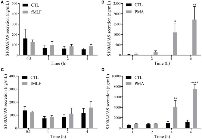
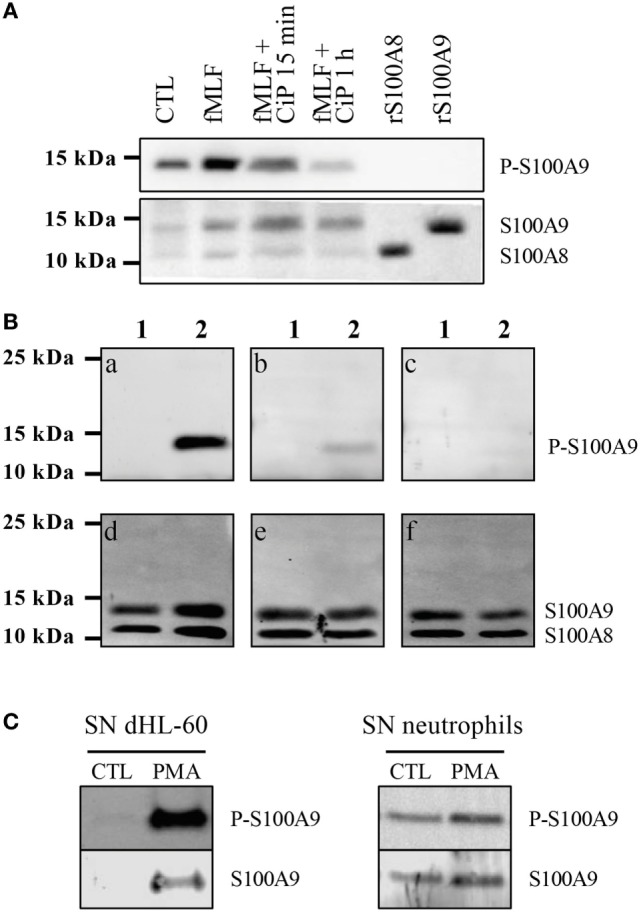
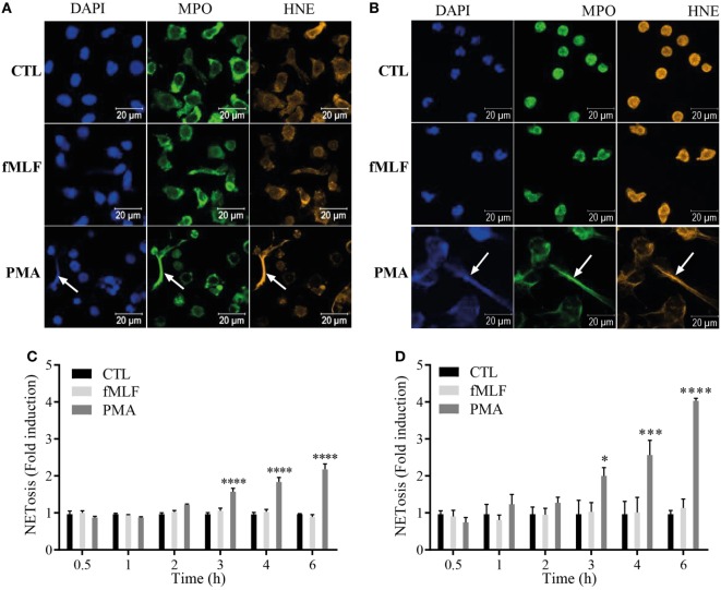
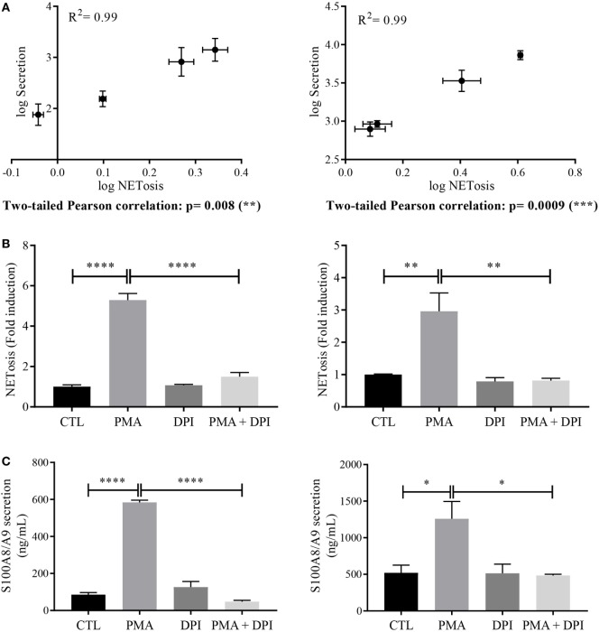
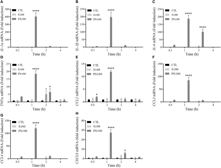
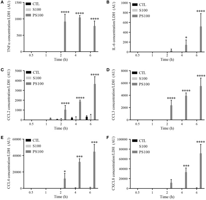
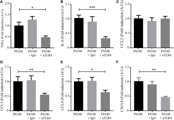
S100A8 and S100A9 are members of the S100 family of cytoplasmic EF-hand Ca2+-binding proteins and are abundantly expressed in the cytosol of neutrophils. In addition to their intracellular roles, S100A8/A9 can be secreted in the extracellular environment and are considered as alarmins able to amplify the inflammatory response. The intracellular activity of S100A8/A9 was shown to be regulated by S100A9 phosphorylation, but the importance of this phosphorylation on the extracellular activity of S100A8/A9 has not yet been extensively studied. Our work focuses on the impact of the phosphorylation state of secreted S100A9 on the proinflammatory function of neutrophils. In a first step, we characterized the secretion of S100A8/A9 in different stimulatory conditions and investigated the phosphorylation state of secreted S100A9. Our results on neutrophil-like differentiated HL-60 (dHL-60) cells and purified human neutrophils showed a time-dependent secretion of S100A8/A9 when induced by phorbol 12-myristoyl 13-acetate and this secreted S100A9 was found in a phosphorylated form. Second, we evaluated the impact of this phosphorylation on proinflammatory cytokine expression and secretion in dHL-60 cells. Time course experiments with purified unphosphorylated or phosphorylated S100A8/A9 were performed and the expression and secretion levels of interleukin (IL)-1α, IL-1β, IL-6, tumor necrosis factor alpha, CCL2, CCL3, CCL4, and CXCL8 were measured by real-time PCR and cytometry bead array, respectively. Our results demonstrate that only the phosphorylated form of the complex induces proinflammatory cytokine expression and secretion. For the first time, we provide evidence that S100A8/PhosphoS100A9 is inducing cytokine secretion through toll-like receptor 4 signaling.

Keywords: S100A8/PhosphoA9; Toll-like receptor 4; cytokines; inflammation; neutrophils.


Figures

Figure 1
Figure 1
Secretion profile of S100A8/A9 under fMLF and phorbol 12-myristoyl 13-acetate (PMA) stimulation. Secretion of S100A8/A9 from differentiated HL-60 cells (A,B) or neutrophils (C,D) after stimulation with 100 nM fMLF (A,C) or 100 nM PMA (B,D) at different time points. S100A8/A9 concentrations were measured in cell supernatants by ELISA. Results are presented as mean ± SEM of five independent experiments. *p < 0.05; **p < 0.01; ***p < 0.001; ****p < 0.0001.
Figure 2
Figure 2
S100A9 is secreted under phosphorylated form. Validation of the custom-made phospho-specific S100A9 antibody (A,B). (A) Non-stimulated cell lysates (CTL), fMLF-stimulated (100 nM, 10 min), phosphatase (CiP)-treated fMLF-stimulated cell lysates as well as recombinant S100A8 and S100A9 (2 μg) were run on a Tris-Tricine gel (10% acrylamide), transferred to PVDF membrane and probed with the anti-PhosphoS100A9 antibody and with the Mac387 antibody to detect total S100A8 and S100A9. (B) 3 μg of purified S100A8/A9 (lane 1) and S100A8/PhosphoA9 (lane 2) were run on a Tris-Tricine gel (10% acrylamide), transferred to PVDF membrane and probed with anti-PhosphoS100A9 antibody alone (a), preincubated 1 h with 1 μg/mL (b) or 4 μg/mL (c) of immunogenic peptide. The same membrane was also probed with B-5 and EPR3554 antibody in order to detect total S100A8 and S100A9 (d–f). (C) Supernatants (SNs) of control or PMA-stimulated dHL60 cells or purified neutrophils were concentrated with Centricon® devices (cut-off 3 kDa) and then loaded on a 10% Tris-Tricine Gel. Western blots were probed with the anti-PhosphoS100A9 antibody and total S100A9 was detected by the Mac387 antibody.
Figure 3
Figure 3
S100A8/A9 are secreted by NETosis from both differentiated HL-60 (dHL-60) cells and purified neutrophils. (A,B) Immunofluorescent staining of neutrophil extracellular traps (NETs) from dHL-60 cells (A) or neutrophils (B). dHL-60 cells or neutrophils were stimulated with 100 nM fMLF (middle panels) or 100 nM phorbol 12-myristoyl 13-acetate (PMA) (lower panels) for 4 h and stained for DNA (DAPI; blue staining), myeloperoxidase (MPO; green staining) or neutrophil elastase (HNE; orange staining). Pictures are representative of at least three independent experiments. (C,D) Quantification of NET release from dHL60 cells (C) or neutrophils (D). Extracellular NET-DNA was quantified with the cell-impermeable fluorescent dye Sytox blue and NET-release was detected via fluorescence emission. NET formation was normalized to the non-stimulated control and expressed as fold induction. Results are presented as mean ± SEM of four independent experiments. *p < 0.05; **p < 0.01; ***p < 0.001; ****p < 0.0001.
Figure 4
Figure 4
Relation between S100 secretion and neutrophil extracellular trap (NET) formation. (A) Correlation between NETosis and S100A8/A9 secretion. Log values of S100A8/A9 secretion were plotted against log values of NETosis and a two-tailed Pearson correlation test was performed for differentiated HL-60 (dHL-60) cells (left panel) or neutrophils (right panel). Involvement of nicotinamide adenine dinucleotide phosphate-oxidase (NADPH oxidase) in NETosis (B) and S100A8/A9 secretion (C) dHL-60 cells (left panel) or neutrophils (right panel) were incubated for 30 min with diphenyleneiodonium (10 μM) then stimulated with phorbol 12-myristoyl 13-acetate (PMA) (100 nM) for 6 h. S100A8/A9 concentration and extracellular NET-DNA were determined in the same condition than in Figures 1 and 3, respectively. Results are presented as mean ± SEM of three (for dHL60) or four (for neutrophils) independent experiments. *p < 0.05; **p < 0.01; ***p < 0.001; ****p < 0.0001.
Figure 5
Figure 5
Cytokine mRNA expression after S100A8/A9 or S100A8/PhosphoA9 stimulation of differentiated HL-60 (dHL-60) cells. dHL-60 cells were stimulated for 30 min, 1, 2, and 4 h with 3 μg/mL of either S100A8/A9 or S100A8/PhosphoA9. Expression of interleukin (IL)-1α (A), IL-1β (B), IL-6 (C), tumor necrosis factor alpha (TNFα) (D), CCL2 (E), CCL3 (F), CCL4 (G), and CXCL8 (H) mRNA were measured by real-time PCR. Results were normalized to three reference genes (Actin-β, B2M, and Gusβ) according to the method of Vandesompele et al. (31) and expressed as fold induction compared to the non-stimulated control (22). Results are presented as mean ± SEM of three independent experiments. *p < 0.05; **p < 0.01; ***p < 0.001; ****p < 0.0001.
Figure 6
Figure 6
Time-dependent cytokine secretion from differentiated HL-60 cells after S100A8/A9 or S100A8/PhosphoA9 stimulation. Cytokine secretion was measured by cytometric bead array upon stimulation with 3 μg/mL S100A8/A9 or S100A8/PhosphoS100A9 for 30 min to 6 h. Tumor necrosis factor alpha (TNFα) (A), interleukin (IL)-6 (B), CCL2 (C), CCL3 (D), CCL4 (E), or CXCL8 (F) concentrations were divided by LDH values in order to correct for cytokine release by cell death. Results are presented as mean ± SEM of three independent experiments. *p < 0.05; **p < 0.01; ***p < 0.001; ****p < 0.0001.
Figure 7
Figure 7
TLR4-dependence of S100A8/PhosphoA9-induced cytokine secretion. Differentiated HL-60 cells were incubated with 1 μg/mL of neutralizing toll-like receptor 4 (TLR4) antibody (aTLR4) or isotype control (IgG) and stimulated with 3 μg/mL S100A8/PhosphoA9 (PS100) for 4 h. Cytokine secretion was measured by cytometric bead array. Concentrations for tumor necrosis factor alpha (TNFα) (A), interleukin (IL)-6 (B), CCL2 (C), CCL3 (D), CCL4 (E), or CXCL8 (F) were divided by LDH values and the results expressed as fold induction of the S100A8/PhosphoA9-stimulated control. Results are presented as mean ± SEM of five independent experiments. *p < 0.05; **p < 0.01; ***p < 0.001; ****p < 0.0001.
Figure 8
Figure 8
Proposed model for the proinflammatory response induced by S100A8/PhosphoA9. Neutrophils activated under different stimulatory conditions (PMA, MSU, Aspergillus fumigates, or Aspergillus nidulans) are releasing NETs through a mechanism involving NADPH oxidase, PAD4, NE, and MPO (34). During NET formation, active S100A8/A9 are released in the extracellular space in a phosphorylated form. S100A8/PhosphoA9 can then activate surrounding neutrophils to secrete cytokines (TNFα and IL-6) and chemokines (CCL2, CCL3, CCL4, and CXCL8). This release is mainly regulated through TLR4 signaling pathways, although other receptors (such as RAGE) are involved in S100A8/PhosphoA9-mediated CCL2 secretion. Thus, S100A8/PhosphoA9 released from neutrophils are involved in the amplification of the inflammatory process and could be the hallmark of various inflammatory diseases. MPO, myeloperoxidase; MSU, monosodium urate monohydrate; NE, neutrophil elastase; PAD4, peptidyl arginine deiminase; PMA, phorbol 12-myristoyl 13-acetate; NET, neutrophil extracellular trap; NADPH oxidase, nicotinamide adenine dinucleotide phosphate-oxidase; TNFα, tumor necrosis factor alpha; IL-6, interleukin 6.

References

    1. Chan JK, Roth J, Oppenheim JJ, Tracey KJ, Vogl T, Feldmann M, et al. Alarmins: awaiting a clinical response. J Clin Invest (2012) 122:2711–9. 10.1172/JCI62423 - DOI - PMC - PubMed
    1. Vogl T, Tenbrock K, Ludwig S, Leukert N, Ehrhardt C, van Zoelen MA, et al. Mrp8 and Mrp14 are endogenous activators of toll-like receptor 4, promoting lethal, endotoxin-induced shock. Nat Med (2007) 13:1042–9. 10.1038/nm1638 - DOI - PubMed
    1. Ehrchen JM, Sunderkotter C, Foell D, Vogl T, Roth J. The endoge-nous toll-like receptor 4 agonist S100A8/S100A9 (calprotectin) as innate amplifier of infection, autoimmunity, and cancer. J Leukoc Biol (2009) 86:557–66. 10.1189/jlb.1008647 - DOI - PubMed
    1. Berthier S, Paclet MH, Lerouge S, Roux F, Vergnaud S, Coleman AW, et al. Changing the conformation state of cytochrome b558 initiates NADPH oxidase activation: MRP8/MRP14 regulation. J Biol Chem (2003) 278:25499–508. 10.1074/jbc.M209755200 - DOI - PubMed
    1. Schenten V, Brechard S, Plancon S, Melchior C, Frippiat JP, Tschirhart EJ. iPLA2, a novel determinant in Ca2+- and phosphorylation-dependent S100A8/A9 regulated NOX2 activity. Biochim Biophys Acta (2010) 1803:840–7. 10.1016/j.bbamcr.2010.02.006 - DOI - PubMed

Publication types

LinkOut - more resources