. 2018 Mar;14(2):396-406.
doi: 10.5114/aoms.2016.63260. Epub 2016 Oct 26.

Preventive effects of total saponins of Panax japonicus on fatty liver fibrosis in mice

Affiliations

Preventive effects of total saponins of Panax japonicus on fatty liver fibrosis in mice

Ding Yuan et al. Arch Med Sci. 2018 Mar.

Abstract

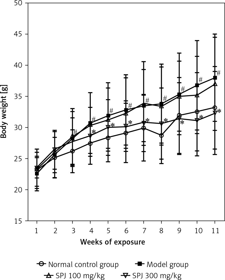
Introduction: Nonalcoholic fatty liver disease (NAFLD) is a condition in which excess fat accumulates in the liver of a patient without a history of alcohol abuse. Fatty liver fibrosis, a severe form of NAFLD, is a key step which can be reversed by effective medical intervention. This paper aims to describe the protective role and mechanisms of action of total saponins of Panax japonicus (SPJ) against fatty liver fibrosis in mice. In this study, fatty liver fibrosis was induced by a high-fat (HF) diet combined with intraperitoneal injection of porcine serum.

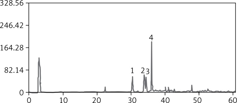
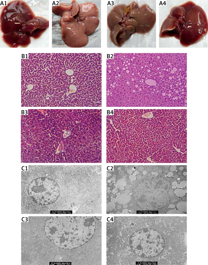
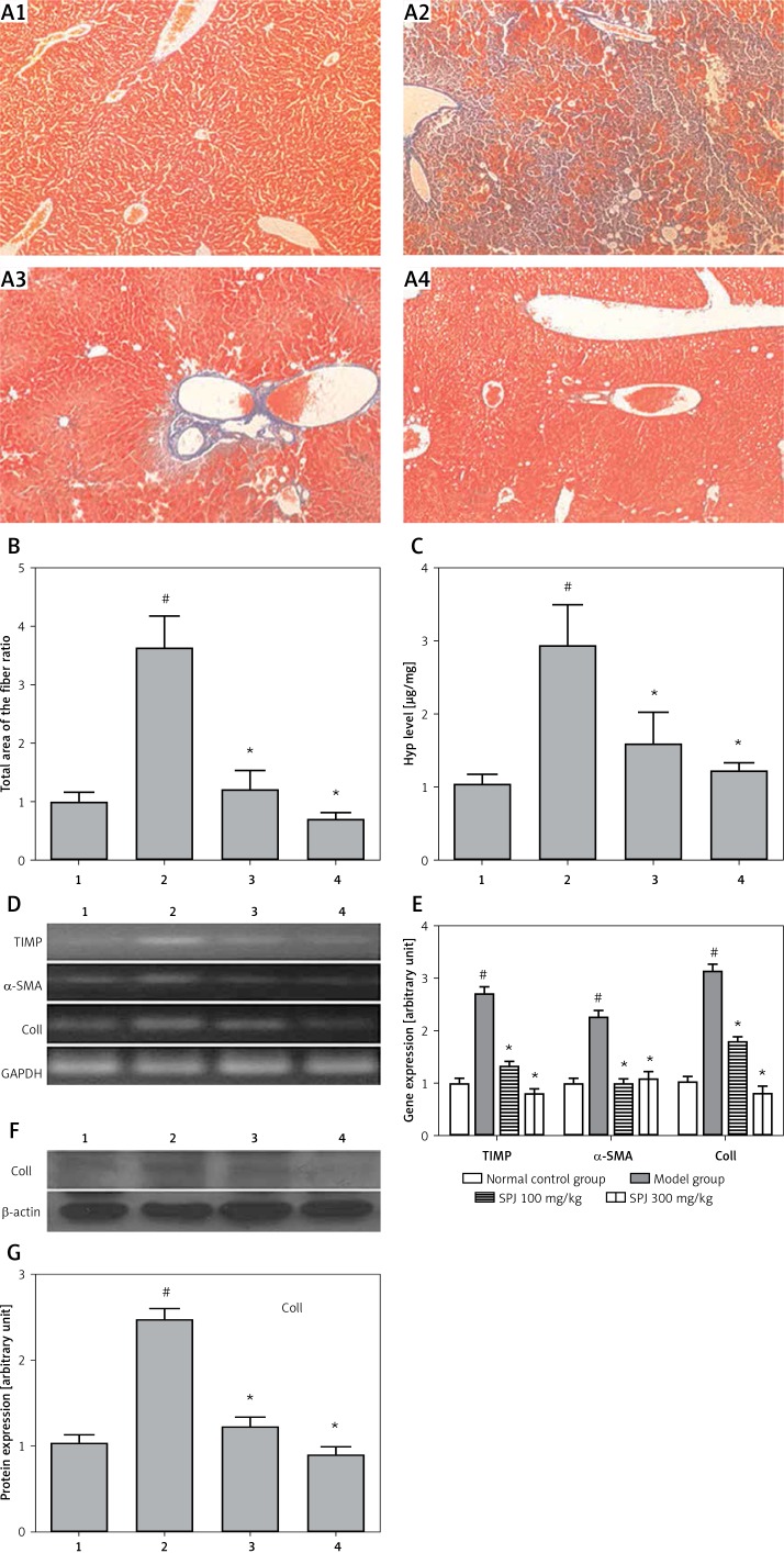
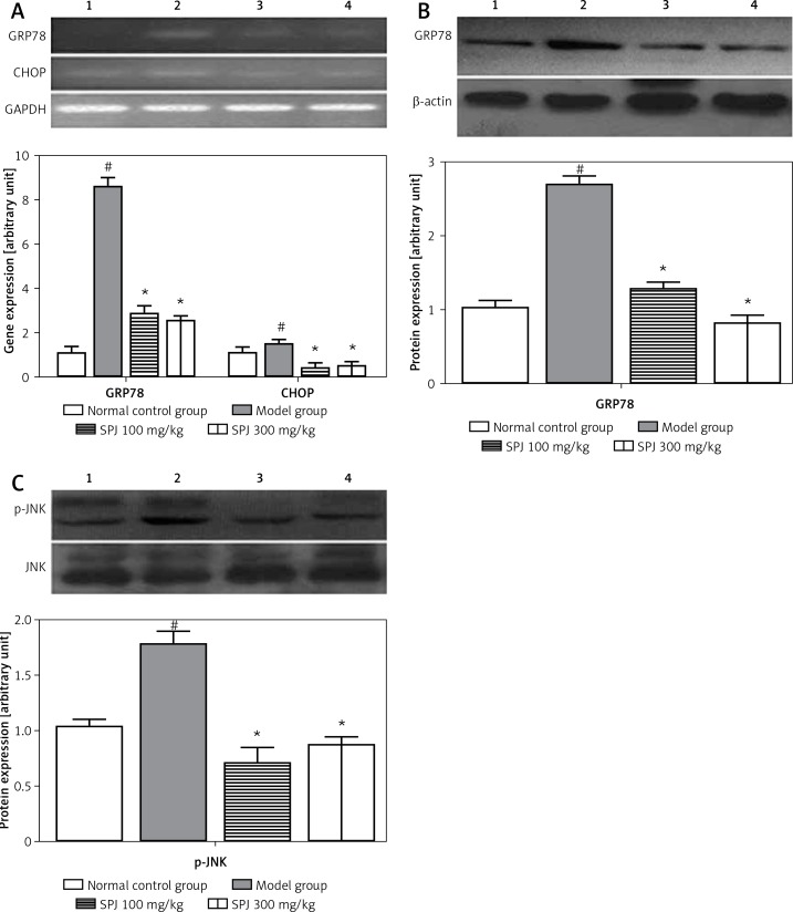
Material and methods: The fatty liver fibrosis model was induced by HF diet combined with intraperitoneal injection of porcine serum. The endoplasmic reticulum stress (ERS) response and C/EBP homologous protein (CHOP) and p-Jun N-terminal kinase (JNK)-mediated apoptosis and inflammation were assessed by serum biochemistry, hematoxylin-eosin (H + E), Masson and electronic microscopy staining, Hyp content detection, Western blotting and real time polymerase chain reaction (RT-PCR).

Results: Saponins of Panax japonicus could significantly improve liver function and decrease the lipid level in the serum. The liver steatosis, collagen fibers and inflammatory cell infiltration were significantly improved in the SPJ group according to microscope observation. The RT-PCR analysis revealed that the collagen I (Coll), α smooth muscle actin (α-SMA), tissue inhibitors of MMPs (TIMP), CHOP and GRP78 mRNA expression levels were distinctly weakened by SPJ treatment; and western blotting analysis indicated that the phosphorylated JNK (p-JNK), Coll and 78 kD glucose-regulated protein (GRP78) protein expression levels were significantly alleviated, which might be associated with the inhibition of the ERS response and the CHOP and JNK-mediated apoptosis and inflammation pathway.

Conclusions: Based on this research, SPJ as a preventive medicine has great potential in prevention of liver fibrosis.

Keywords: Coll; JNK; endoplasmic reticulum stress; fatty liver fibrosis; total saponins of Panax japonicus.


Conflict of interest statement

The authors declare no conflict of interest.

Figures

Figure 1
Figure 1
The saponins of Panax japonicus analyzed by HPLC-ELSD 1 – chikusetsusaponin V, 2 – Pjs-2, 3 – chikusetsusaponin IV, 4 – chikusetsusaponin IVa.
Figure 2
Figure 2
Changes of mice body weight. The data of body weight are presented as means ± SD (n = 6) #p < 0.01 vs. the normal control group; *p < 0.01 vs. the model group.
Figure 3
Figure 3
Pathological features of liver tissue. A – Morphologic changes in liver: (1) normal control group, liver appeared fresh, slippery and dense both in color and texture; (2) model group, on liver surface there appeared numerous tiny milky white particles; liver had rough appearance and grey color; (3) SPJ low dose group (100 mg/kg) and (4) SPJ high dose group (300 mg/kg), the liver appearance was improved dramatically, especially in the SPJ high dose group; the liver was redder, smoother and denser. B – H + E staining of liver tissue: (1) normal control group, showing a normal morphological structure in lobular architecture and hepatocytes; (2) model group, revealing conspicuous morphological disruption; (3) SPJ low dose group (100 mg/kg), displaying moderate improvement of morphological changes; (4) SPJ high dose group (300 mg/kg), demonstrating obvious improvement in the hepatocytes. Liver sections showed a dose-dependent reduction in the degree of liver injury. C – Ultrastructural observation of the hepatocytes: (1) normal control group, the cell ultrastructure was quite regular including mitochondria and endoplasmic reticulum; (2) model group, showing numerous fat vacuoles in the hepatocytes and accompanied by a large area of damage and fracture of the mitochondria and the mitochondrial crest; (3) SPJ low dose group (100 mg/kg), revealing some mitochondria and fewer fat vacuoles in the hepatocytes; (4) SPJ high dose group (300 mg/kg), revealing almost normal mitochondria and endoplasmic reticulum in the cellular ultrastructure 1 – normal control group, 2 – model group, 3 – SPJ 100 mg/kg, 4 – SPJ 300 mg/kg.
Figure 4
Figure 4
SPJ treatment inhibited collagen deposition in the mouse liver. A – Masson’s trichrome staining of liver tissues (magnification, 200×): (1) normal control group, normal structure of the hepatic lobule; (2) model group, extensive accumulation of continuous fibrotic septa was around the central vein in the liver; (3) SPJ low dose group (100 mg/kg), showing less collagen deposited and pseudolobule formation; (4) SPJ high dose group (300 mg/kg), collagen fiber rarely existed. B – Relative quantitative analysis of the fibrosis in Masson staining: using software analyses the fiber sensitivity, in the fatty liver fibrosis group, the sum area of the fiber was raised evidently; SPJ (100 mg/kg and 300 mg/kg) groups showed a distinctly smaller sum area of the fiber; the sum area of the fiber ratio is presented as means ± SD (n = 6). #p < 0.01 vs. normal control group; *p < 0.01 vs. model group. C – Hyp content detection in the liver tissue: compared with the model group, the Hyp content sharply declined with the SPJ treatment (100 mg/kg and 300 mg/kg); the Hyp content is presented as means ± SD (n = 6). #p < 0.01 vs. normal control group; *p < 0.01 vs. model group. D and E – Effect of SPJ on the expression of TIMP, α-SMA, COLL Iα in the liver tissues: SPJ treatment (100 mg/kg and 300 mg/kg) reduced TIMP, α-SMA, COLL Iα gene expression compared with liver fibrosis group; however, in the SPJ high dose group (300 mg/kg) the decrease was more obvious; the gene expression levels are presented as means ± SD (n = 6). #p < 0.01 vs. normal control group; *p < 0.01 vs. model group. F and G – Expression of COLL Iα protein in the liver tissues: with SPJ treatment especially the high dose group (300 mg/kg) significantly downregulated levels of COLL Iα compared with the model group; the protein expression levels are presented as means ± SD (n = 6). #p < 0.01 vs. normal control group; *p < 0.01 vs. model group 1 – normal control group, 2 – model group, 3 – SPJ 100 mg/kg, 4 – SPJ 300 mg/kg.
Figure 5
Figure 5
SPJ treatment recovered the expression of ERS-associated genes and proteins. A – Effects of SPJ on the gene expression of GRP78 and CHOP in the liver tissues: in SPJ (100 mg/kg and 300 mg/kg), the mRNA levels of GRP78 and CHOP were remarkably decreased compared with the model group, the gene expression levels are presented as means ± SD (n = 6). #p < 0.01 vs. normal control group; *p < 0.01 vs. model group. B – Effects of SPJ on protein expression of GRP78 in the liver tissues: anti-β-actin blotting was used as control for equal protein loading. Compared with the model group, SPJ (100 mg/kg and 300 mg/kg) treatment significantly suppressed the protein expression of GRP78; the protein expression levels are presented as means ± SD (n = 6). #p < 0.01 vs. normal control group; *p < 0.01 vs. model group. C – Effects of SPJ on the protein expression of JNK and p-JNK in the liver tissues: Noticeable changes of the JNK protein expression did not appear in the normal control group, model group and SPJ (100 mg/kg and 300 mg/kg) groups, while the expression of p-JNK was significantly decreased in SPJ (100 mg/kg and 300 mg/kg) groups in contrast with the model group; protein expression levels are presented as means ±SD (n = 6). #p < 0.01 vs. normal control group; *p < 0.01 vs. model group 1 – normal control group, 2 – model group, 3 – SPJ 100 mg/kg, 4 – SPJ 300 mg/kg.

Similar articles

Cited by

References

    1. Dyson J, Day C. Treatment of non-alcoholic fatty liver disease. Dig Dis. 2014;32:597–604. - PubMed
    1. Paschos P, Paletas K. Non alcoholic fatty liver disease and metabolic syndrome. Hippokratia. 2009;13:9–19. - PMC - PubMed
    1. Lu Y, Wei J, Tang Y, et al. Evaluation of fatty liver fibrosis in rabbits using real-time shear wave elastography. Exp Ther Med. 2014;8:355–62. - PMC - PubMed
    1. Balunas MJ, Kinghorn AD. Drug discovery from medicinal plants. Life Sci. 2005;78:431–41. - PubMed
    1. He YM, Lu KM, Yuan D, Zhang CC. Studies on preparative technology and quantitative determination for extracts of total saponin in roof of Panax japonicus. Zhongguo Zhong Yao Za Zhi. 2008;33:2607–11. - PubMed