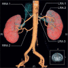
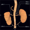
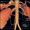
Morphological and clinical aspects of the occurrence of accessory (multiple) renal arteries
- PMID: 29593819
- PMCID: PMC5868651
- DOI: 10.5114/aoms.2015.55203
Morphological and clinical aspects of the occurrence of accessory (multiple) renal arteries
Abstract
Renal vascularization variants vastly differ between individuals due to the very complex embryogenesis of the kidneys. Moreover, each variant may have implications for clinical and surgical interventions. The number of operating procedures continues to grow, and includes renal transplants, aneurysmorrhaphy and other vascular reconstructions. In any surgical technique, unawareness of the presence of multiple renal arteries may result in a fatal outcome, especially if laparoscopic methods are used. The aim of this review is to comprehensively identify the variation within multiple renal arteries and to highlight the connections between the presence of accessory renal arteries and the coexistence of other variants of vascularization. Another aim is to determine the potential clinical implications of the presence of accessory renal arteries. This study is of particular importance for surgeons, intervention radiologists, nephrologists and vascular surgeons.
Keywords: accessory; anatomical variation; kidney; multiple; renal artery.
Conflict of interest statement
The authors declare no conflict of interest.
Figures


Similar articles
-
Accessory (multiple) renal arteries - Differences in frequency according to population, visualizing techniques and stage of morphological development.Vascular. 2016 Oct;24(5):531-7. doi: 10.1177/1708538116631223. Epub 2016 Mar 4. Vascular. 2016. PMID: 26945775 Review.
-
Frequencies of accessory renal arteries in 129 Iranian patients.Am J Clin Exp Urol. 2020 Feb 25;8(1):38-42. eCollection 2020. Am J Clin Exp Urol. 2020. PMID: 32211452 Free PMC article.
-
A right ectopic kidney with bilateral multiple anomalies of the renal vasculature - a case report.J Clin Diagn Res. 2013 Jan;7(1):150-3. doi: 10.7860/JCDR/2012/5000.2692. Epub 2012 Oct 20. J Clin Diagn Res. 2013. PMID: 23450664 Free PMC article.
-
Does the type of renal artery anatomic variant determine the diameter of the main vessel supplying a kidney? A study based on CT data with a particular focus on the presence of multiple renal arteries.Surg Radiol Anat. 2018 Apr;40(4):381-388. doi: 10.1007/s00276-017-1930-z. Epub 2017 Oct 5. Surg Radiol Anat. 2018. PMID: 28980056 Free PMC article.
-
Short- and long-term outcomes of kidney transplants with multiple renal arteries.Ann Surg. 1995 Apr;221(4):406-14. doi: 10.1097/00000658-199504000-00012. Ann Surg. 1995. PMID: 7726677 Free PMC article. Review.
Cited by
-
A Unique Case of Incomplete Bifid Ureter and Associated Arterial Variations.Case Rep Urol. 2021 Jan 4;2021:6655813. doi: 10.1155/2021/6655813. eCollection 2021. Case Rep Urol. 2021. PMID: 33489410 Free PMC article.
-
Anatomic Variations of Renal Arteries as an Important Factor in the Effectiveness of Renal Denervation in Resistant Hypertension.J Cardiovasc Dev Dis. 2023 Aug 29;10(9):371. doi: 10.3390/jcdd10090371. J Cardiovasc Dev Dis. 2023. PMID: 37754800 Free PMC article. Review.
-
Image-guided study of swine anatomy as a tool for urologic surgery research and training.Acta Cir Bras. 2021 Jan 20;35(12):e351208. doi: 10.1590/ACB351208. eCollection 2021. Acta Cir Bras. 2021. PMID: 33503221 Free PMC article.
-
SCAI Position Statement on Renal Denervation for Hypertension: Patient Selection, Operator Competence, Training and Techniques, and Organizational Recommendations.J Soc Cardiovasc Angiogr Interv. 2023 Aug 21;2(6Part A):101121. doi: 10.1016/j.jscai.2023.101121. eCollection 2023 Nov-Dec. J Soc Cardiovasc Angiogr Interv. 2023. PMID: 39129887 Free PMC article. No abstract available.
-
Wunderlich Syndrome: Spontaneous Cystic Rupture on Account of Acquired Kidney Atrophy.Cureus. 2022 Oct 17;14(10):e30386. doi: 10.7759/cureus.30386. eCollection 2022 Oct. Cureus. 2022. PMID: 36407245 Free PMC article.
References
-
- Satyapal KS, Haffejee AA, Singh B, Ramsaroop L, Robbs JV, Kalideen JM. Additional renal arteries: incidence and morphometry. Surg Radiol Anat. 2001;23:33–8. - PubMed
-
- Singh D, Finelli A, Rubinstein M, Desai MM, Kaouk J, Gill IS. Laparoscopic partial nephrectomy in the presence of multiple renal arteries. Urology. 2007;69:444–7. - PubMed
-
- Eustachi Opuscula anatomia. Venice: p. 1564.
-
- Merklin RJ, Michels NA. The variant renal and suprarenal blood supply with data on the inferior phrenic, ureteral and gonadal arteries: a statistical analysis based on 185 dissections and reviews of the literature. J Int Coll Surg. 1958;29:41–76. - PubMed
LinkOut - more resources
Full Text Sources
Other Literature Sources
