

Role of Mast Cells in Oral Lichen Planus and Oral Lichenoid Reactions
- PMID: 29593898
- PMCID: PMC5822832
- DOI: 10.1155/2018/7936564
Role of Mast Cells in Oral Lichen Planus and Oral Lichenoid Reactions
Abstract
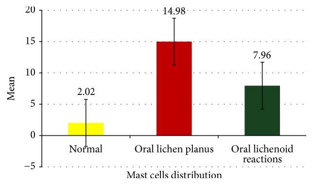
Introduction: Oral lichen planus (OLP) is a chronic T cell mediated disease of oral mucosa, skin, and its appendages with a prevalence of 0.5 to 2.6% worldwide. Oral lichenoid reactions (OLR) are a group of lesions with diverse aetiologies but have clinical and histological features similar to OLP, thereby posing a great challenge in differentiating both lesions. Mast cells are multifunctional immune cells that play a major role in the pathogenesis of lichen planus by release of certain chemical mediators. Increased mast cell densities with significant percentage of degranulation have been observed as a consistent finding in pathogenesis of oral lichen planus.
Aim: The current study was aimed at quantifying the mast cells in histopathological sections of OLP and OLR thereby aiding a means of distinguishing these lesions.
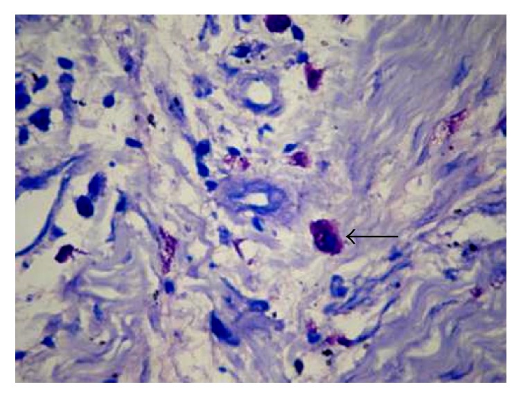
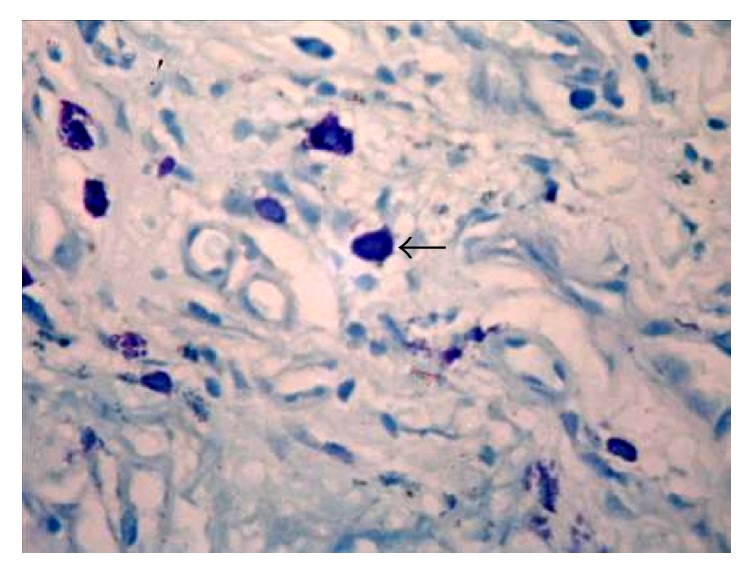
Materials and methods: The study group involved 21 cases of oral lichen planus, 21 cases of oral lichenoid reactions, and 10 control specimens of normal buccal mucosa. All the cases were stained with Toluidine Blue and routine haematoxylin and eosin and the mast cells were quantified.
Statistical analysis used: The results were analyzed using the Kruskal-Wallis test and an intergroup analysis was performed using Mann-Whitney U test.
Conclusion: The number of mast cells showed an increased value in oral lichen planus when compared to oral lichenoid reaction and thus an estimation of mast cells count could aid in distinguishing OLP from OLR histopathologically.
Figures
Similar articles
-
To Investigate the Involvement of Mast Cells in the Pathogenesis of Oral Lichen Planus and Oral Lichenoid Reactions.J Pharm Bioallied Sci. 2024 Dec;16(Suppl 5):S4755-S4759. doi: 10.4103/jpbs.jpbs_913_24. Epub 2025 Jan 30. J Pharm Bioallied Sci. 2024. PMID: 40061733 Free PMC article.
-
Role of mast cells in pathogenesis of oral lichen planus.J Oral Maxillofac Pathol. 2011 Sep;15(3):267-71. doi: 10.4103/0973-029X.86674. J Oral Maxillofac Pathol. 2011. PMID: 22144827 Free PMC article.
-
A histochemical and immunohistochemical study of mast cells in differentiating oral lichen planus from oral lichenoid reactions.Quintessence Int. 2010 Mar;41(3):221-7. Quintessence Int. 2010. PMID: 20213023
-
Oral lichen planus and lichenoid reactions: etiopathogenesis, diagnosis, management and malignant transformation.J Oral Sci. 2007 Jun;49(2):89-106. doi: 10.2334/josnusd.49.89. J Oral Sci. 2007. PMID: 17634721 Review.
-
ORAL LICHEN PLANUS AND ORAL LICHENOID REACTION--AN UPDATE.Acta Clin Croat. 2015 Dec;54(4):516-20. Acta Clin Croat. 2015. PMID: 27017728 Review.
Cited by
-
Correlation of Mast Cell and Angiogenesis in Oral Lichen Planus, Dysplasia (Leukoplakia), and Oral Squamous Cell Carcinoma.Rambam Maimonides Med J. 2021 Apr 29;12(2):e0016. doi: 10.5041/RMMJ.10438. Rambam Maimonides Med J. 2021. PMID: 33938803 Free PMC article.
-
Comprehensive Insight into Lichen Planus Immunopathogenesis.Int J Mol Sci. 2023 Feb 3;24(3):3038. doi: 10.3390/ijms24033038. Int J Mol Sci. 2023. PMID: 36769361 Free PMC article. Review.
-
Association of Mast Cell Activity With Chronic Gingivitis, Chronic Periodontitis, and Aggressive Periodontitis in Adults: A Histochemical Observational Study.Cureus. 2025 Jun 25;17(6):e86762. doi: 10.7759/cureus.86762. eCollection 2025 Jun. Cureus. 2025. PMID: 40718240 Free PMC article.
-
Lichen Planus: What is New in Diagnosis and Treatment?Am J Clin Dermatol. 2024 Sep;25(5):735-764. doi: 10.1007/s40257-024-00878-9. Epub 2024 Jul 9. Am J Clin Dermatol. 2024. PMID: 38982032 Review.
-
Mast cells in oral lichen planus and oral lichenoid lesions related to dental amalgam contact.Braz Oral Res. 2024 Jan 5;38:e005. doi: 10.1590/1807-3107bor-2024.vol38.0005. eCollection 2024. Braz Oral Res. 2024. PMID: 38198305 Free PMC article.
References
-
- Shirasuna K. Oral lichen planus: Malignant potential and diagnosis. Oral Science International. 2014;11(1):1–7. doi: 10.1016/S1348-8643(13)00030-X. - DOI
-
- van der Meij E. H., van der Waal I. Lack of clinicopathologic correlation in the diagnosis of oral lichen planus based on the presently available diagnostic criteria and suggestions for modifications. Journal of Oral Pathology & Medicine. 2003;32(9):507–512. doi: 10.1034/j.1600-0714.2003.00125.x. - DOI - PubMed
LinkOut - more resources
Full Text Sources
Other Literature Sources
Medical
