

Enhanced Photodynamic Cancer Treatment by Mitochondria-Targeting and Brominated Near-Infrared Fluorophores
- PMID: 29593951
- PMCID: PMC5867131
- DOI: 10.1002/advs.201700481
Enhanced Photodynamic Cancer Treatment by Mitochondria-Targeting and Brominated Near-Infrared Fluorophores
Abstract
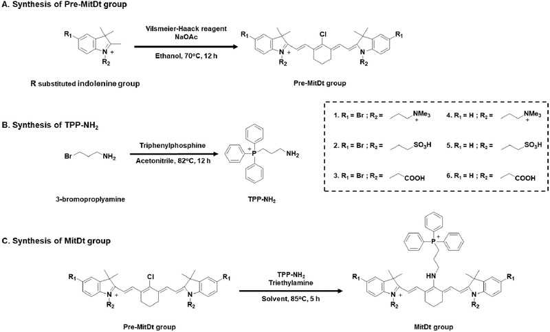
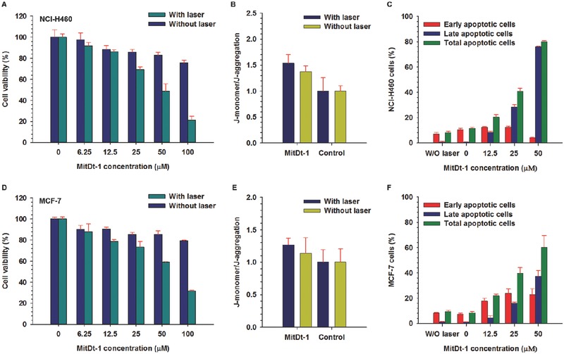
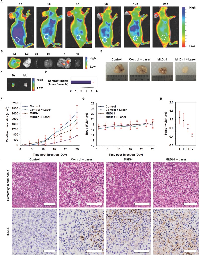
A noninvasive and selective therapy, photodynamic therapy (PDT) is widely researched in clinical fields; however, the lower efficiency of PDT can induce unexpected side effects. Mitochondria are extensively researched as target sites to maximize PDT effects because they play crucial roles in metabolism and can be used as cancer markers due to their high transmembrane potential. Here, a mitochondria targeting photodynamic therapeutic agent (MitDt) is developed. This photosensitizer is synthesized from heptamethine cyanine dyes, which are conjugated or modified as follows. The heptamethine meso-position is conjugated with a triphenylphosphonium derivative for mitochondrial targeting, the N-alkyl side chain is modified for regulation of charge balance and solubility, and the indolenine groups are brominated to enhance reactive oxygen species generation (ROS) after laser irradiation. The synthesized MitDt increases the cancer uptake efficiency due to the lipo-cationic properties of the triphenylphosphonium, and the PDT effects of MitDt are amplified after laser irradiation because mitochondria are susceptible to ROS, the response to which triggers an apoptotic anticancer effect. Consequently, these hypotheses are demonstrated by in vitro and in vivo studies, and the results indicate strong potential for use of MitDts as efficient single-molecule-based PDT agents for cancer treatment.
Keywords: cancer therapy; heptamethine cyanine dye; mitochondria targeting; near‐infrared (NIR) dye; photodynamic therapy.
Figures
Similar articles
-
Synthesis of a versatile mitochondria-targeting small molecule for cancer near-infrared fluorescent imaging and radio/photodynamic/photothermal synergistic therapies.Mater Today Bio. 2022 Jun 7;15:100316. doi: 10.1016/j.mtbio.2022.100316. eCollection 2022 Jun. Mater Today Bio. 2022. PMID: 35721281 Free PMC article.
-
The investigation of unique water-soluble heptamethine cyanine dye for use as NIR photosensitizer in photodynamic therapy of cancer cells.Spectrochim Acta A Mol Biomol Spectrosc. 2020 Mar 5;228:117702. doi: 10.1016/j.saa.2019.117702. Epub 2019 Nov 1. Spectrochim Acta A Mol Biomol Spectrosc. 2020. PMID: 31748160
-
A Selenium-Substituted Heptamethine Cyanine Photosensitizer for Near-Infrared Photodynamic Therapy.Chembiochem. 2022 Nov 18;23(22):e202200421. doi: 10.1002/cbic.202200421. Epub 2022 Oct 18. Chembiochem. 2022. PMID: 36149045
-
Potential of Cyanine Derived Dyes in Photodynamic Therapy.Pharmaceutics. 2021 May 31;13(6):818. doi: 10.3390/pharmaceutics13060818. Pharmaceutics. 2021. PMID: 34072719 Free PMC article. Review.
-
Recent progress on near-infrared fluorescence heptamethine cyanine dye-based molecules and nanoparticles for tumor imaging and treatment.Wiley Interdiscip Rev Nanomed Nanobiotechnol. 2023 Sep-Oct;15(5):e1910. doi: 10.1002/wnan.1910. Epub 2023 Jun 12. Wiley Interdiscip Rev Nanomed Nanobiotechnol. 2023. PMID: 37305979 Review.
Cited by
-
Mitochondrial Targeting and Imaging with Small Organic Conjugated Fluorophores: A Review.Chemistry. 2022 Dec 27;28(72):e202202366. doi: 10.1002/chem.202202366. Epub 2022 Oct 26. Chemistry. 2022. PMID: 36121738 Free PMC article. Review.
-
Site-selected thionated benzothioxanthene chromophores as heavy-atom-free small-molecule photosensitizers for photodynamic therapy.Commun Chem. 2022 Oct 31;5(1):142. doi: 10.1038/s42004-022-00752-x. Commun Chem. 2022. PMID: 36697939 Free PMC article.
-
Brain endothelial cell-derived extracellular vesicles with a mitochondria-targeting photosensitizer effectively treat glioblastoma by hijacking the blood‒brain barrier.Acta Pharm Sin B. 2023 Sep;13(9):3834-3848. doi: 10.1016/j.apsb.2023.03.023. Epub 2023 Mar 31. Acta Pharm Sin B. 2023. PMID: 37719366 Free PMC article.
-
Cyanine dyes in the mitochondria-targeting photodynamic and photothermal therapy.Commun Chem. 2024 Aug 13;7(1):180. doi: 10.1038/s42004-024-01256-6. Commun Chem. 2024. PMID: 39138299 Free PMC article. Review.
-
X-ray induced photodynamic therapy (PDT) with a mitochondria-targeted liposome delivery system.J Nanobiotechnology. 2020 Jun 10;18(1):87. doi: 10.1186/s12951-020-00644-z. J Nanobiotechnology. 2020. PMID: 32522291 Free PMC article.
References
-
- Dolmans D. E., Fukumura D., Jain R. K., Nat. Rev. Cancer 2003, 3, 380. - PubMed
-
- Noh I., Kim H. O., Choi J., Choi Y., Lee D. K., Huh Y. M., Haam S., Biomaterials 2015, 53, 763. - PubMed
-
- a) Konopka K., Goslinski T., J. Dent. Res. 2007, 86, 694; - PubMed
- b) Nomoto T., Fukushima S., Kumagai M., Miyazaki K., Inoue A., Mi P., Maeda Y., Toh K., Matsumoto Y., Morimoto Y., Kishimura A., Nishiyama N., Kataoka K., Biomater. Sci. 2016, 4, 826; - PubMed
- c) Yoo J., Lee D., Gujrati V., Rejinold N. S., Lekshmi K. M., Uthaman S., Jeong C., Park I. K., Jon S., Kim Y. C., J. Controlled Release 2016, 28, 142. - PubMed
LinkOut - more resources
Full Text Sources
Other Literature Sources
Miscellaneous