Review
. 2018 Feb;7(1):70-82.
doi: 10.21037/tau.2017.12.31.

MRI in early prostate cancer detection: how to manage indeterminate or equivocal PI-RADS 3 lesions?

Affiliations
Review

MRI in early prostate cancer detection: how to manage indeterminate or equivocal PI-RADS 3 lesions?

Ivo G Schoots. Transl Androl Urol. 2018 Feb.

Abstract

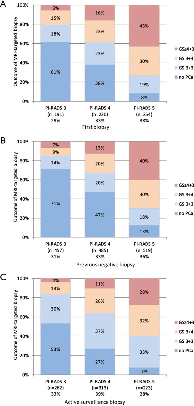
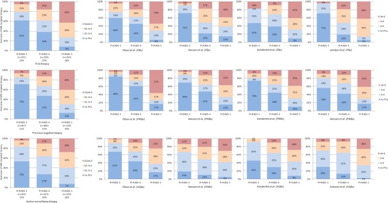
This review focuses on indeterminate lesions on prostate magnetic resonance imaging (MRI), assigned as PI-RADS category 3. The prevalence of PI-RADS 3 index lesion in the diagnostic work-up is significant, varying between one in three (32%) to one in five (22%) men, depending on patient cohort of first biopsies, previously negative biopsies, and active surveillance biopsies. A management strategy must be developed for this group of men with an indeterminate suspicion of having clinically significant prostate cancer (csPCa). Currently available data show that the actual prevalence of csPCa after targeted biopsy in PI-RADS 3 lesions vary between patients groups from one in five (21%) to one in six (16%), depending on previous biopsy status. Although this prevalence is lower in comparison to PI-RADS 4 and PI-RADS 5 lesions, still a considerable proportion of men harbor significant disease. Men with such a PI-RADS 3 lesion should therefore be adequately managed. In general, the clinical approach of using a threshold of PI-RADS ≥4 instead of PI-RADS ≥3 to select MRI for targeted biopsies is not supported by data from our explorative literature search using current definitions of csPCa. A possible adaptation to the threshold of PI-RADS ≥4 in combination with other clinical markers could be considered within an active surveillance protocol, where the balance between the individual risk of missing csPCa and the constant process of repeating prostate biopsies is crucial. In the future, improvements in MR imaging and interpretation, combined with molecular biomarkers and multivariate risk models will all be employed in prostate cancer detection and monitoring. These combinations will aid decision-making in challenging circumstances, such as unclear and diagnostic equivocal results for csPCa at early detection.

Keywords: MRI-guided targeted biopsy; PI-RADS; PI-RADS 3; PSA density; Prostate cancer; biopsy; equivocal; indeterminate; magnetic resonance imaging (MRI); risk stratification.


Conflict of interest statement

Conflicts of Interest: The author has no conflicts of interest to declare.

Figures

Figure 1
Figure 1
A left peripheral zone lesion (red arrows) on (A) axial T2-weighted image, (B) high-b-value DWI, (C) early DCE time-point, and (D) ADC map. The overall PI-RADS assessment category 3 was assigned (T2w 3, DWI/ADC 3, DCE −). On subsequent targeted MRI/US fusion biopsy, the lesion exhibited Gleason score 3+4 tumor (red arrows). PI-RADS category 3 lesions are characterized as focal mildly to moderately hypointense on ADC, and isointense to mildly hyperintense on high b-value DWI. This is in combination with heterogeneous signal intensity or non-circumscribed, rounded, and moderate hypointensity on T2w images. No focal enhancement on DCE MRI should be visualized. The dominant MRI sequence in PI-RADS v2 is DWI/ADC. (E-H) A left peripheral zone lesion (blue arrows). The overall PI-RADS assessment category 3 was assigned (T2w 3, DWI/ADC 3, DCE −). On subsequent targeted MRI/US fusion biopsy, the lesion exhibited benign prostatic tissue (blue arrows).
Figure 2
Figure 2
A left transition zone lesion (arrow) on (A) axial T2-weighted image, (B) high-b-value DWI, (C) early DCE time-point, and (D) ADC map. The overall PI-RADS assessment category 3 was assigned (T2w 3, DWI/ADC 4, DCE +). On subsequent targeted MRI/US fusion biopsy, the lesion exhibited Gleason score 3+4 tumor (red arrows). PI-RADS category 3 lesions in the transition zone are characterized as heterogeneous signal intensity with obscured margins, together with focal mildly to moderately hypointense on ADC, and isointense to mildly hyperintense on high b-value DWI. Focal enhancement on DCE MRI may be present. The dominant sequence in PI-RADS v2 is T2w. (E-H) A left transition zone lesion (blue arrows). The overall PI-RADS assessment category 3 was assigned (T2w 3, DWI/ADC 4, DCE −). On subsequent targeted MRI/US fusion biopsy, the lesion exhibited no prostate cancer (blue arrows).
Figure 3
Figure 3
Histology outcome by Gleason score (GS) of PI-RADS assessment categories 3, 4 and 5, with subsequently MRI-targeted biopsies, in men with first biopsies (A), previous negative biopsies (B), and active surveillance biopsies (C). Cumulated data, generated by individual reports (7,8,10,12,13,21) (see Figure S1).
Figure S1
Figure S1
Histology outcome by Gleason score (GS) of PI-RADS assessment categories 3, 4 and 5, with subsequently MRI-targeted biopsies, in men with first biopsies, previous negative biopsies, and active surveillance biopsies. Cumulated data (left), generated by individual reports (middle to right) (7,8,10,12,13,21).

Similar articles

Cited by

References

    1. Mottet N, Bellmunt J, Bolla M, et al. EAU-ESTRO-SIOG Guidelines on Prostate Cancer. Part 1: Screening, Diagnosis, and Local Treatment with Curative Intent. Eur Urol 2017;71:618-29. 10.1016/j.eururo.2016.08.003 - DOI - PubMed
    1. Moldovan PC, Van den Broeck T, Sylvester R, et al. What Is the Negative Predictive Value of Multiparametric Magnetic Resonance Imaging in Excluding Prostate Cancer at Biopsy? A Systematic Review and Meta-analysis from the European Association of Urology Prostate Cancer Guidelines Panel. Eur Urol 2017;72:250-66. 10.1016/j.eururo.2017.02.026 - DOI - PubMed
    1. Dickinson L, Ahmed HU, Allen C, et al. Magnetic resonance imaging for the detection, localisation, and characterisation of prostate cancer: recommendations from a European consensus meeting. Eur Urol 2011;59:477-94. 10.1016/j.eururo.2010.12.009 - DOI - PubMed
    1. Barentsz JO, Richenberg J, Clements R, et al. ESUR prostate MR guidelines 2012. Eur Radiol 2012;22:746-57. 10.1007/s00330-011-2377-y - DOI - PMC - PubMed
    1. Weinreb JC, Barentsz JO, Choyke PL, et al. PI-RADS Prostate Imaging - Reporting and Data System: 2015, Version 2. Eur Urol 2016;69:16-40. 10.1016/j.eururo.2015.08.052 - DOI - PMC - PubMed