. 2018 Jan 30;2(3):207-219.
doi: 10.1210/js.2017-00338. eCollection 2018 Mar 1.

Enhanced Expression of the Key Mitosis Regulator Cyclin B1 Is Mediated by PDZ-Binding Kinase in Islets of Pregnant Mice

Affiliations

Enhanced Expression of the Key Mitosis Regulator Cyclin B1 Is Mediated by PDZ-Binding Kinase in Islets of Pregnant Mice

Tadayoshi Uesato et al. J Endocr Soc. .

Abstract

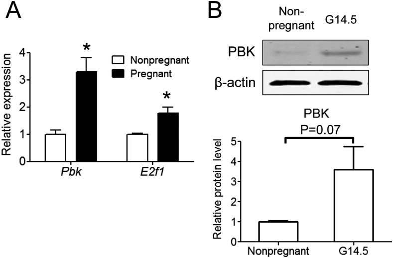
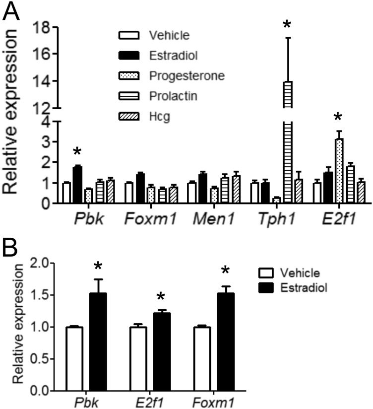
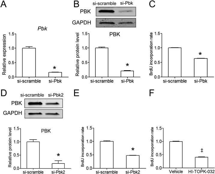
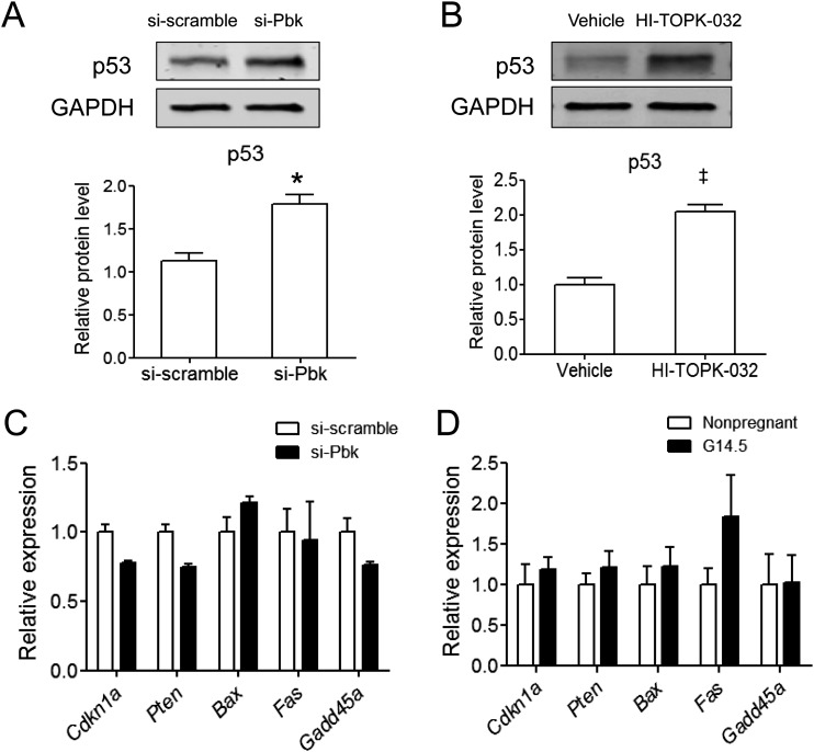
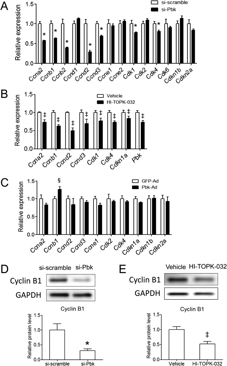
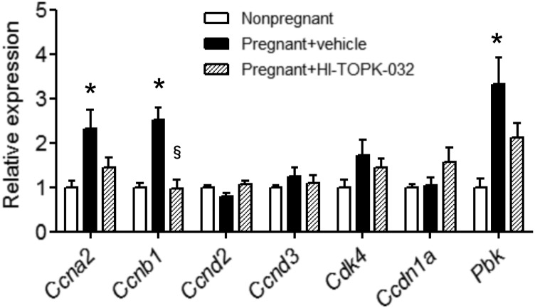
The proliferation of pancreatic β cells is enhanced to enable an increase in β-cell mass and to compensate for insulin resistance during pregnancy. To elucidate the mechanisms involved, we previously investigated islets from pregnant and nonpregnant mice by gene expression profiling and found that the expression of postsynaptic density-95/Discs large/zonula occludens-1 (PDZ)-binding kinase (Pbk), a member of the mitogen-activated protein kinase kinase family, is increased in pregnant mouse islets compared with control mouse islets. Among the pregnancy hormones, treatment with estradiol upregulated Pbk expression. Inhibition of Pbk expression using a small interfering RNA for Pbk reduced bromodeoxyuridine incorporation in mouse insulinoma 6 cells, which was accompanied by a decreased expression of Ccnb1, a regulatory gene involved in mitosis. Ccnb1 expression was augmented in mouse islets during pregnancy. The forced expression of Pbk using an adenovirus system in isolated mouse islets increased Ccnb1 expression, and the Pbk inhibitor HI-TOPK-032 suppressed Ccnb1 expression in islets isolated from pregnant mice. Our results suggest that Pbk contributes to the expansion of islets during pregnancy and that Ccnb1 may assist Pbk in its role in β-cell proliferation.

Keywords: Pbk; cyclin B1; estradiol; islet; p53.


Conflict of interest statement

T.O. has received research funding from AstraZeneca, Eli Lilly and Company, Sanofi-Aventis, and Mitsubishi Tanabe Pharma. The remaining authors have nothing to disclose.

Figures

Figure 1.
Figure 1.
Pbk expression is increased in mouse islets during pregnancy. (A) Islets were isolated from nonpregnant and pregnant mice at G14.5 for qRT-PCR analysis. Expression levels of Pbk and E2f1 were normalized to Tbp expression levels. Results are shown as the means ± standard error of the mean; n ≥ 3. *Statistically significant difference (P < 0.05) compared with the islets isolated from nonpregnant mice. (B) Islets were isolated from nonpregnant and pregnant mice at G14.5 and subjected to immunoblot using PBK and β-actin antibodies. Upper panels show a representative immunoblot. Relative protein levels of Pbk were normalized to β-actin (n = 4).
Figure 2.
Figure 2.
Estradiol upregulates Pbk expression in isolated mouse islets and MIN6 cells. (A) Islets were isolated from 12-week-old female mice; treated with 100 nM estradiol, 10 μM progesterone, 44.2 mM prolactin, or 10 IU/mL human chorionic gonadotropin (Hcg) for 24 hours; and then subjected to qRT-PCR (n ≥ 4). *Statistically significant difference (P < 0.05) compared with islets treated with vehicle. (B) MIN6 cells were treated with or without 100 nM estradiol for 24 hours. Total RNA was isolated for qRT-PCR analysis (n ≥ 4). Expression levels of each gene were normalized to Tbp expression. *Statistically significant difference (P < 0.05) compared with MIN6 cells treated with vehicle. Results are shown as the means ± standard error of the mean.
Figure 3.
Figure 3.
Pbk knockdown reduces cell proliferation in mouse insulinoma MIN6 cells. (A) Seventy-two hours after transfection with si-Pbk or si-scramble, total RNA was isolated from MIN6 cells for analysis of Pbk expression. Expression levels of Pbk were normalized to Tbp expression (n ≥ 3). (B) After knockdown using si-Pbk or si-scramble, total protein was isolated from MIN6 cells and subjected to immunoblot for PBK and GAPDH. Upper panels show a representative immunoblot. Relative protein levels of Pbk were normalized to Gapdh (n ≥ 3). (C) Seventy-two hours after the transfection of MIN6 cells with si-Pbk or si-scramble, the BrdU incorporation assay was performed. (D) Seventy-two hours after transfection with si-Pbk2 or si-scramble, total protein was isolated from MIN6 cells and subjected to immunoblot for PBK and GAPDH. (E) After knockdown using si-Pbk2 or si-scramble, MIN6 cells were subjected to BrdU incorporation analysis. (F) Twenty-four hours after treatment with HI-TOPK-032 (5 μM) or vehicle, BrdU incorporation analysis was performed. Results are shown as the mean ± standard error of the mean. *Statistically significant difference (P < 0.05) compared with MIN6 cells treated with si-scramble. ‡Statistically significant difference (P < 0.05) compared with MIN6 cells treated with vehicle.
Figure 4.
Figure 4.
Expression levels of p53-target genes are not altered by Pbk knockdown in MIN6 cells. (A) Seventy-two hours after transfection of MIN6 cells with si-Pbk or si-scramble, total protein was isolated for immunoblot analysis. Upper panels show representative immunoblots of p53 and GAPDH. Relative protein levels of p53 were normalized to those of Gapdh (n ≥ 4). *Statistically significant difference (P < 0.05) compared with MIN6 cells transfected with si-scramble. (B) Six hours after treatment with HI-TOPK-032 (5 μM) or vehicle, total protein was isolated for immunoblot analysis using anit-p53 and GAPDH antibodies. ‡Statistically significant difference (P < 0.05) compared with MIN6 cells treated with vehicle. (C) After knockdown using si-Pbk or si-scramble, total RNA was isolated from MIN6 cells to analyze the expression levels of p53-target genes, including Cdkn1a, Pten, Bax, Fas, and Gadd45a. Expression levels of each gene were normalized to those of Tbp. Results are shown as the means ± standard error of the mean; n ≥ 3. (D) Total RNA was isolated from islets of pregnant or nonpregnant mice to analyze the expression levels of p53-target genes (n ≥ 4).
Figure 5.
Figure 5.
Pbk knockdown attenuates Ccnb1 expression in MIN6 cells. (A) Expression levels of genes regulating the cell cycle were analyzed by qRT-PCR in MIN6 cells transfected with si-Pbk or si-scramble. *Statistically significant difference (P < 0.05) compared with MIN6 cells transfected with si-scramble. (B) Six hours after treatment with HI-TOPK-032 (5 μM) or vehicle, total RNA was isolated from MIN6 cells for qRT-PCR analysis (n ≥ 3). ‡Statistically significant difference (P < 0.05) compared with MIN6 cells treated with vehicle. (C) Isolated mouse islets were incubated with adenoviruses expressing Pbk or GFP and then subjected to qRT-PCR analysis (n = 3). Expression levels of each gene were normalized to those of Tbp. Results are shown as the mean ± standard error of the mean. §Statistically significant difference (P < 0.05) compared with islets treated with green fluorescent protein–expressing adenoviruses. (D) MIN6 cells were transfected with si-Pbk or si-scramble, followed by immunoblotting for PBK and GAPDH. Upper panels show representative immunoblots. The lower panel shows relative protein levels of Pbk, which were normalized to those of Gapdh (n ≥ 3). *Statistically significant difference (P < 0.05) compared with MIN6 cells transfected with si-scramble. (E) Six hours after treatment with HI-TOPK-032 or vehicle, total protein was isolated from MIN6 cells for immunoblot analysis. ‡Statistically significant difference (P < 0.05) compared with vehicle-treated cells. Results are shown as the mean ± standard error of the mean; n ≥ 3.
Figure 6.
Figure 6.
Attenuation of Ccnb1 expression by a Pbk inhibitor in isolated islets of pregnant mice. Isolated mouse islets were incubated with 5 μM HI-TOPK-032 or vehicle for 24 hours and then subjected to qRT-PCR analysis. Expression levels of each gene were normalized to Tbp expression (n ≥ 4). *Statistically significant difference (P < 0.05) compared with islets of nonpregnant mice. §Statistically significant difference (P < 0.05) compared with islets of pregnant mice treated with vehicle.

References

    1. Buchanan TA, Xiang AH. Gestational diabetes mellitus. J Clin Invest. 2005;115(3):485–491. - PMC - PubMed
    1. Rieck S, Kaestner KH. Expansion of beta-cell mass in response to pregnancy. Trends Endocrinol Metab. 2010;21(3):151–158. - PMC - PubMed
    1. Huang C, Snider F, Cross JC. Prolactin receptor is required for normal glucose homeostasis and modulation of beta-cell mass during pregnancy. Endocrinology. 2009;150(4):1618–1626. - PubMed
    1. Hughes E, Huang C. Participation of Akt, menin, and p21 in pregnancy-induced beta-cell proliferation. Endocrinology. 2011;152(3):847–855. - PubMed
    1. Karnik SK, Chen H, McLean GW, Heit JJ, Gu X, Zhang AY, Fontaine M, Yen MH, Kim SK. Menin controls growth of pancreatic beta-cells in pregnant mice and promotes gestational diabetes mellitus. Science. 2007;318(5851):806–809. - PubMed

LinkOut - more resources