

Mechanisms of Trabecular Formation and Specification During Cardiogenesis
- PMID: 29594501
- PMCID: PMC6164162
- DOI: 10.1007/s00246-018-1868-x
Mechanisms of Trabecular Formation and Specification During Cardiogenesis
Abstract
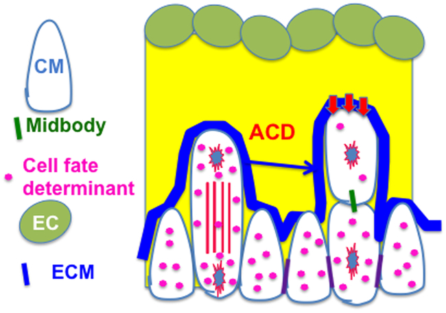
Trabecular morphogenesis is a key morphologic event during cardiogenesis and contributes to the formation of a competent ventricular wall. Lack of trabeculation results in embryonic lethality. The trabecular morphogenesis is a multistep process that includes, but is not limited to, trabecular initiation, proliferation/growth, specification, and compaction. Although a number of signaling molecules have been implicated in regulating trabeculation, the cellular processes underlying mammalian trabecular formation are not fully understood. Recent works show that the myocardium displays polarity, and oriented cell division (OCD) and directional migration of the cardiomyocytes in the monolayer myocardium are required for trabecular initiation and formation. Furthermore, perpendicular OCD is an extrinsic asymmetric cell division that contributes to trabecular specification, and is a mechanism that causes the trabecular cardiomyocytes to be distinct from the cardiomyocytes in compact zone. Once the coronary vasculature system starts to function in the embryonic heart, the trabeculae will coalesce with the compact zone to thicken the heart wall, and abnormal compaction will lead to left ventricular non-compaction (LVNC) and heart failure. There are many reviews about compaction and LVNC. In this review, we will focus on the roles of myocardial polarity and OCD in trabecular initiation, formation, and specification.
Keywords: Myocardial polarity; Oriented cell division; Trabecular specification; Trabeculation.
Conflict of interest statement
Figures
References
-
- Van Mierop LH Embryology of the univentricular heart. Herz 4, 78–85 (1979). - PubMed
-
- Sedmera D, Pexieder T, Vuillemin M, Thompson RP & Anderson RH Developmental patterning of the myocardium. Anat Rec 258, 319–337 (2000). - PubMed
-
- Icardo JM & Fernandez-Teran A Morphologic study of ventricular trabeculation in the embryonic chick heart. Acta Anat (Basel) 130, 264–274 (1987). - PubMed
Publication types
MeSH terms
Grants and funding
LinkOut - more resources
Full Text Sources
Other Literature Sources
