Color Doppler ultrasound diagnosis of intrarenal vein thrombosis: A rare case report and literature review
- PMID: 29595692
- PMCID: PMC5895364
- DOI: 10.1097/MD.0000000000010284
Color Doppler ultrasound diagnosis of intrarenal vein thrombosis: A rare case report and literature review
Abstract
Rationale: We present a case of intrarenal vein thrombosis (IRVT) diagnosed by ultrasound (US). To the best of our knowledge, this is the first reported case in the imaging literature.
Patient concerns: A 15-year-old boy with a 4-year history of thrombocytopenic purpura presented to the emergency room with a 2-day history of sudden-onset severe left flank pain associated with gross hematuria.
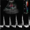
Diagnoses: Hypercholesterolemia, proteinuria, and elevated plasma creatinine level were present. The US examination showed obscurely structured, sparsely distributed arterial and venous flow signals, and an increased resistance index (RI) in a localized area. The diagnosis was acute renal failure and nephrotic syndrome accompanied by left IRVT.
Interventions: The patient was treated with anticoagulation therapy for 1 month.
Outcomes: Clinical symptoms were relieved. The US re-examination revealed that the arterial flow spectra had returned to normal. Also, more venous flow signals were observed in the involved area, suggesting thrombolysis.
Lessons: This previously unreported case should alert sonographers to include IRVT in the differential diagnosis of flank pain associated with hematuria. In such cases, both kidneys and different areas of the same kidney should be scanned and compared. Some features, including an obscure structure and an increased RI for the involved area indicate possible IRVT.
Conflict of interest statement
The authors have no funding and conflicts of interest to disclose.
Figures


References
-
- Decoster T, Schwagten V, Hendriks J, et al. Renal colic as the first symptom of acute renal vein thrombosis, resulting in the diagnosis of nephrotic syndrome. Eur J Emerg Med 2009;16:170–1. - PubMed
-
- Llach F, Papper S, Massry SG. The clinical spectrum of renal vein thrombosis: acute and chronic. Am J Med 1980;69:819–27. - PubMed
-
- Asghar M, Ahmed K, Shah SS, et al. Renal vein thrombosis. Eur J Vasc Endovasc Surg 2007;34:217–23. - PubMed
-
- Zubarev AV. Ultrasound of renal vessels. Eur Radiol 2001;11:1902–15. - PubMed
-
- Gonzalez R, Schwartz S, Sheldon CA, et al. Bilateral renal vein thrombosis in infancy and childhood. Urol Clin North Am 1982;9:279–83. - PubMed
Publication types
MeSH terms
Substances
LinkOut - more resources
Full Text Sources
Other Literature Sources
Medical

