

Rare cardiac metastasis of soft tissue sarcoma: A case report and literature review
- PMID: 29595697
- PMCID: PMC5895417
- DOI: 10.1097/MD.0000000000009814
Rare cardiac metastasis of soft tissue sarcoma: A case report and literature review
Abstract
Rationale: Owing to the unique structure and function of the heart, tumor metastasis in the heart is rare. Accordingly, no unique symptoms have yet been identified for cardiac metastasis.
Patient and concerns: A patient presented with cardiac metastasis 3 years after surgical resection of alveolar soft tissue sarcomas in their late stage.
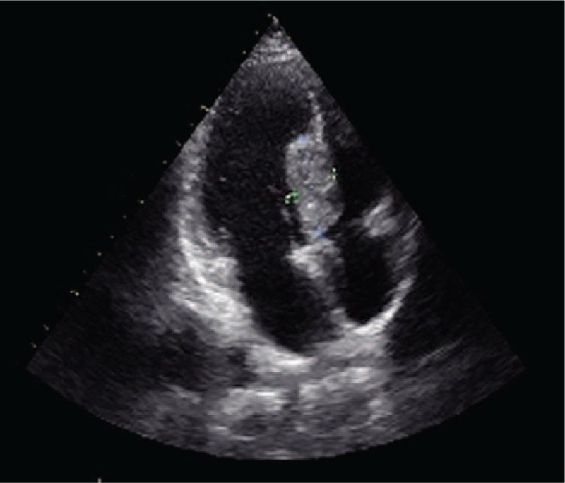
Diagnosis: Ultrasonography showed a middle-high echo clump on the left surface of the mid-upper interventricular septum, which had an unclear boundary with the myocardium. Meanwhile, blood flow was found in the clump, with no blockage of blood flow having been observed.
Lessons: Although cardiac metastasis in terminal cancer patients always carries a poor prognosis, there is still no effective treatment for cardiac metastasis. In the clinic, it is important to improve the patient's quality of life, reduce symptoms and signs, and extend the duration of survival.
Conflict of interest statement
The authors have no funding and conflicts of interest to disclose.
Figures

References
-
- Wang ZJ, Reddy GP, Gotway MB, et al. CT and MR imaging of pericardial disease. Radiographics 2003;23:167–80. - PubMed
-
- Díaz ML, Villanueva A, Bastarrika G, et al. Non-electrocardiogram-gated multidetector-row computed tomography findings of cardiac pathology in oncologic patients. Curr Probl Diagn Radiol 2009;38:206–17. - PubMed
-
- Artioli G, Borgato L, Calamelli S, et al. Unusual cardiac metastasis of uterine leiomyosarcoma: case report and literature review. Tumori 2016;102(Suppl. 2): - PubMed
-
- Gül M, Babat N, Uçar FM, et al. Massive pulmonary embolism and a cardiac mass: thrombus or metastasis? Turk Kardiyol Dern Ars 2016;44:597–9. - PubMed
-
- Haverkamp MC, Scholte AJ, Holman ER, et al. Contrast echocardiography as a useful additional diagnostic tool in evaluating a primary cardiac tumor. Eur J Echocardiogr 2005;6:388–91. - PubMed
Publication types
MeSH terms
LinkOut - more resources
Full Text Sources
Other Literature Sources
Medical
