Imaging Bacterial Cell Wall Biosynthesis
- PMID: 29596002
- PMCID: PMC6287495
- DOI: 10.1146/annurev-biochem-062917-012921
Imaging Bacterial Cell Wall Biosynthesis
Abstract
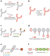
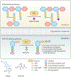
Peptidoglycan is an essential component of the cell wall that protects bacteria from environmental stress. A carefully coordinated biosynthesis of peptidoglycan during cell elongation and division is required for cell viability. This biosynthesis involves sophisticated enzyme machineries that dynamically synthesize, remodel, and degrade peptidoglycan. However, when and where bacteria build peptidoglycan, and how this is coordinated with cell growth, have been long-standing questions in the field. The improvement of microscopy techniques has provided powerful approaches to study peptidoglycan biosynthesis with high spatiotemporal resolution. Recent development of molecular probes further accelerated the growth of the field, which has advanced our knowledge of peptidoglycan biosynthesis dynamics and mechanisms. Here, we review the technologies for imaging the bacterial cell wall and its biosynthesis activity. We focus on the applications of fluorescent d-amino acids, a newly developed type of probe, to visualize and study peptidoglycan synthesis and dynamics, and we provide direction for prospective research.
Keywords: bacterial cell wall; bacterial morphogenesis; d-amino acids; fluorescent probes; microscopy; peptidoglycan.
Figures


Similar articles
-
d-Amino Acid Derivatives as in Situ Probes for Visualizing Bacterial Peptidoglycan Biosynthesis.Acc Chem Res. 2019 Sep 17;52(9):2713-2722. doi: 10.1021/acs.accounts.9b00311. Epub 2019 Aug 16. Acc Chem Res. 2019. PMID: 31419110 Review.
-
Super-resolution microscopy reveals cell wall dynamics and peptidoglycan architecture in ovococcal bacteria.Mol Microbiol. 2011 Dec;82(5):1096-109. doi: 10.1111/j.1365-2958.2011.07871.x. Epub 2011 Nov 7. Mol Microbiol. 2011. PMID: 22059678
-
In Situ probing of newly synthesized peptidoglycan in live bacteria with fluorescent D-amino acids.Angew Chem Int Ed Engl. 2012 Dec 7;51(50):12519-23. doi: 10.1002/anie.201206749. Epub 2012 Oct 10. Angew Chem Int Ed Engl. 2012. PMID: 23055266 Free PMC article.
-
New chemical tools to probe cell wall biosynthesis in bacteria.Curr Opin Microbiol. 2015 Oct;27:69-77. doi: 10.1016/j.mib.2015.07.013. Epub 2015 Aug 25. Curr Opin Microbiol. 2015. PMID: 26291270 Review.
-
Fluorescent d-Amino Acids for Super-resolution Microscopy of the Bacterial Cell Wall.ACS Chem Biol. 2022 Sep 16;17(9):2418-2424. doi: 10.1021/acschembio.2c00496. Epub 2022 Aug 22. ACS Chem Biol. 2022. PMID: 35994360
Cited by
-
Discovery of MurA Inhibitors as Novel Antimicrobials through an Integrated Computational and Experimental Approach.Antibiotics (Basel). 2022 Apr 14;11(4):528. doi: 10.3390/antibiotics11040528. Antibiotics (Basel). 2022. PMID: 35453279 Free PMC article.
-
Chemical Reporters for Exploring Microbiology and Microbiota Mechanisms.Chembiochem. 2020 Jan 15;21(1-2):19-32. doi: 10.1002/cbic.201900535. Epub 2019 Dec 27. Chembiochem. 2020. PMID: 31730246 Free PMC article. Review.
-
Carbohydrate-based drugs launched during 2000-2021.Acta Pharm Sin B. 2022 Oct;12(10):3783-3821. doi: 10.1016/j.apsb.2022.05.020. Epub 2022 May 23. Acta Pharm Sin B. 2022. PMID: 36213536 Free PMC article. Review.
-
Super-Resolution Microscopy of the Bacterial Cell Wall Labeled by Fluorescent D-Amino Acids.Methods Mol Biol. 2024;2727:83-94. doi: 10.1007/978-1-0716-3491-2_7. Methods Mol Biol. 2024. PMID: 37815710
-
Large-Scale Discovery of Microbial Fibrillar Adhesins and Identification of Novel Members of Adhesive Domain Families.J Bacteriol. 2022 Jun 21;204(6):e0010722. doi: 10.1128/jb.00107-22. Epub 2022 May 24. J Bacteriol. 2022. PMID: 35608365 Free PMC article.
References
-
- CDC (Cent. Dis. Control Prev.). 2013. Antibiotic resistance threats in the United States, 2013 US Dept. Health Hum. Serv., Washington, DC
-
- Vollmer W, Blanot D, de Pedro MA. 2008. Peptidoglycan structure and architecture. FEMS Microbiol. Rev 32(2):149–67 - PubMed
Publication types
MeSH terms
Substances
Grants and funding
LinkOut - more resources
Full Text Sources
Other Literature Sources

