Review
. 2018 Mar 29;19(4):1019.
doi: 10.3390/ijms19041019.

EP4 as a Therapeutic Target for Aggressive Human Breast Cancer

Affiliations
Review

EP4 as a Therapeutic Target for Aggressive Human Breast Cancer

Mousumi Majumder et al. Int J Mol Sci. .

Abstract

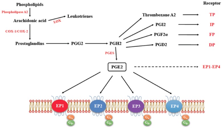
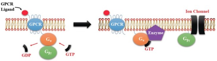
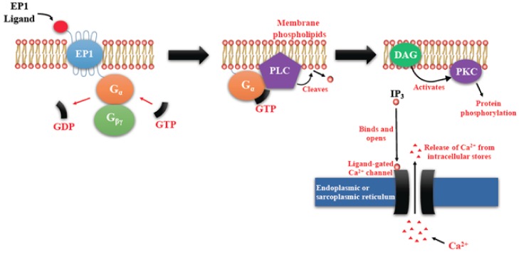
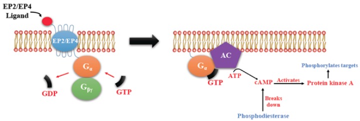
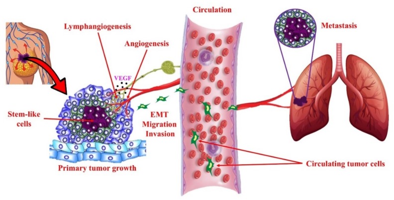
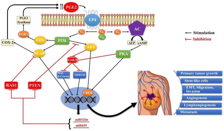
G-protein-coupled receptors (GPCRs, also called seven-transmembrane or heptahelical receptors) are a superfamily of cell surface receptor proteins that bind to many extracellular ligands and transmit signals to an intracellular guanine nucleotide-binding protein (G-protein). When a ligand binds, the receptor activates the attached G-protein by causing the exchange of Guanosine-5'-triphosphate (GTP) for guanosine diphosphate (GDP). They play a major role in many physiological functions, as well as in the pathology of many diseases, including cancer progression and metastasis. Only a few GPCR members have been exploited as targets for developing drugs with therapeutic benefit in cancer. Present review briefly summarizes the signaling pathways utilized by the EP (prostaglandin E receptor) family of GPCR, their physiological and pathological roles in carcinogenesis, with special emphasis on the roles of EP4 in breast cancer progression. We make a case for EP4 as a promising newer therapeutic target for treating breast cancer. We show that an aberrant over-expression of cyclooxygenase (COX)-2, which is an inflammation-associated enzyme, occurring in 40-50% of breast cancer patients leads to tumor progression and metastasis due to multiple cellular events resulting from an increased prostaglandin (PG) E2 production in the tumor milieu. They include inactivation of host anti-tumor immune cells, such as Natural Killer (NK) and T cells, increased immuno-suppressor function of tumor-associated macrophages, promotion of tumor cell migration, invasiveness and tumor-associated angiogenesis, due to upregulation of multiple angiogenic factors including Vascular Endothelial Growth Factor (VEGF)-A, increased lymphangiogenesis (due to upregulation of VEGF-C/D), and a stimulation of stem-like cell (SLC) phenotype in cancer cells. All of these events were primarily mediated by activation of the Prostaglandin (PG) E receptor EP4 on tumor or host cells. We show that selective EP4 antagonists (EP4A) could mitigate all of these events tested with cells in vitro as well as in vivo in syngeneic COX-2 expressing mammary cancer bearing mice or immune-deficient mice bearing COX-2 over-expressing human breast cancer xenografts. We suggest that EP4A can avoid thrombo-embolic side effects of long term use of COX-2 inhibitors by sparing cardio-protective roles of PGI2 via IP receptor activation or PGE2 via EP3 receptor activation. Furthermore, we identified two COX-2/EP4 induced oncogenic and SLC-stimulating microRNAs-miR526b and miR655, one of which (miR655) appears to be a potential blood biomarker in breast cancer patients for monitoring SLC-ablative therapies, such as with EP4A. We suggest that EP4A will likely produce the highest benefit in aggressive breast cancers, such as COX-2 expressing triple-negative breast cancers, when combined with other newer agents, such as inhibitors of programmed cell death (PD)-1 or PD-L1.

Keywords: COX-2; EP receptors; PGE2; angiogenesis; breast cancer; lymphangiogenesis; metastasis; microRNAs; stem-like cells; triple-negative breast cancer.


Conflict of interest statement

The authors declare no conflict of interest.

Figures

Figure 1
Figure 1
The pathway for the synthesis of prostaglandins, their respective receptors and signaling. (Adapted with kind permission from Markovič, T.; et al. 2017; reference [9]). Arachidonic acid acts as the substrate for cyclooxygenase (COX)-1 and COX-2 to produce Prostaglandins PGE2, Thromboxane A2, PGI2, PGF2α, and PGD2, all of which exert functions by binding to their respective receptors. EP1-EP4 (dashed arrow) receptors are further detailed for their G protein coupling.
Figure 2
Figure 2
Heterotrimeric G-protein activation and inactivation cycle. The activation occurs by conversion of G-protein alpha (Gα)-coupled guanosine diphosphate (GDP) to Guanosine-5′-triphosphate (GTP). The activated G-protein then dissociates into an α and a β/γ complex. GTP bound Gα is active. Intrinsic GTPase activity leads to the inactivation of the G-Protein. GDP bound Gα re-associates with a β/γ complex to form the inactive G-protein that can again associate with a receptor.
Figure 3
Figure 3
Prostaglandin E receptors (EP1)-mediated signaling events. EP1 couples with Gq, activating PLC that cleaves PIP2, to generate second messengers, IP3, and diacylglycerol (DAG). IP3 binds to and opens a ligand-gated Ca2+ channel in the endoplasmic reticulum leading to an increase in cytosolic Ca2+. Ca2+ in the cytosol exerts its effects by binding to Ca2+-binding proteins.
Figure 4
Figure 4
Shared pathway of EP2/EP4 mediated Signaling. There is activation of adenylyl cyclase (AC) leading to a rise in the second messenger cAMP in the cytosol that activates Protein kinase A (PKA). PKA in turn activates a transcription factor CREB (cAMP response element-binding protein).
Figure 5
Figure 5
EP4 mediated signaling (in addition to PKA activation) not shared by EP2 (adapted with kind permission from O’callaghan, G.; Houston, A.; 2015; reference [13]). There is non-canonical activation of the PI3K-Akt and ERK pathways. Akt, also called protein kinase B (PKB) promotes cell survival by activating the transcription factor NF-κB. ERK is primarily a promoter of cell proliferation and migration. Cell proliferation depends on the ERK mediated activation of the transcription factor EGR-1.
Figure 6
Figure 6
EP3 mediated signaling (adapted with kind permission from O’callaghan, G.; Houston, A.; 2015; reference [13]). EP3 has multiple isoforms, most of which are coupled with the inhibitory G-protein Gi that acts by inhibiting AC-cAMP-PKA pathway. Those coupled with Gs stimulate AC-cAMP-PKA pathway. Those coupled with G12/13 are involved in Rho family GTPase signaling utilized in cell migration by cytoskeleton remodeling.
Figure 7
Figure 7
Schema of cellular events in tumor progression and metastasis. Primary tumor growth depends on proliferation of tumorigenic cells, some of which adopt a stem-like cell (SLC) phenotype under the influence of genetic and epigenetic (micro-environmental) mechanisms. Local tumor growth is dependent on angiogenesis (formation of new blood vessels), which also facilitates tumor cell egress into the circulation. In addition, many epithelial tumors undergo intra-tumoral and/or peri-tumoral lymphangiogenesis (formation of new lymphatic vessels) that helps tumor cells to migrate to lymph nodes and then enter circulation. Epithelial-mesenchymal transition (EMT) is a phenotypic change in epithelial tumor cells utilized for invasion and migration out of the local confines. These cellular events are stimulated in COX-2 expressing breast tumors by activation of EP4 on tumor cells and tumor-associated host cells (immune cells, endothelial cells), so that EP4 presents as a therapeutic target to block multiple cellular events in tumor progression.
Figure 8
Figure 8
Schema of EP4 mediated signaling pathways in COX-2 expressing breast cancer. Aberrant COX- 2 activity leads to tumor progression and metastasis by utilizing multiple signaling pathways in which EP4 activation plays a pivotal role, and two COX-2/EP4 upregulated miRNAs (miR526b and miR655) are important partners. EP4 activation (like EP2) results in cAMP-dependent PKA activation leading to phosphorylation of the transcription factor CREB. PKA also upregulates WNT/β-catenin and NOTCH pathways by inhibiting GSK3. Furthermore, unlike EP2, EP4 also utilizes the non-canonical PI3K/AkT and ERK signaling pathways, respectively promoting cell survival and migration/ proliferation. COX-2 upregulates the miRNAs miR526b and miR655 via EP4 mediated PI3K/Akt activation and WNT/β-catenin/NOTCH pathways. While COX-2 induces these miRNAs, the miRNAs, in turn, upregulated COX-2. We suggest that these occur via upregulation of NF-κB, which is a well-known upregulator of COX-2 under certain conditions. Predicted targets of these miRNAs include NF-κB repressor genes. Thus there appears to exist a positive feedback loop for COX-2/EP4/NF-κB/miRNA/COX-2-mediated SLC perpetuation.

Similar articles

Cited by

References

    1. Lappano R., Maggiolini M. G-protein-coupled receptors: Novel targets for drug discovery in cancer. Nat. Rev. Drug Discov. 2011;10:47–60. doi: 10.1038/nrd3320. - DOI - PubMed
    1. Liu Y., An S., Ward R., Yang Y., Guo X., Li W., Xu T. G-protein-coupled receptors as promising cancer targets. Cancer Lett. 2016;376:226–239. doi: 10.1016/j.canlet.2016.03.031. - DOI - PubMed
    1. Cho S., Jeon J., Kim S.I. Personalized medicine in breast cancer: A systematic review. J. Breast Cancer. 2012;15:265–272. doi: 10.4048/jbc.2012.15.3.265. - DOI - PMC - PubMed
    1. Fan C., Oh D.S., Wessels L., Weigelt B., Nuyten D.S., Nobel A.B., Van’t Veer L.J., Perou C.M. Concordance among gene-expression—Based predictors for breast cancer. N. Engl. J. Med. 2006;355:560–569. doi: 10.1056/NEJMoa052933. - DOI - PubMed
    1. Dai X., Li T., Bai Z., Yang Y., Liu X., Zhan J., Shi B. Breast cancer intrinsic subtype classification, clinical use and future trends. Am. J. Cancer Res. 2015;5:2929–2943. - PMC - PubMed

MeSH terms