. 2018 Mar 29;19(4):1026.
doi: 10.3390/ijms19041026.

Treatment of Arsenite Intoxication-Induced Peripheral Vasculopathy with Mesenchymal Stem Cells

Affiliations

Treatment of Arsenite Intoxication-Induced Peripheral Vasculopathy with Mesenchymal Stem Cells

Yi-Hung Chiang et al. Int J Mol Sci. .

Abstract

Arsenite (As), a notorious toxic metal, is ubiquitously distributed in the earth and poses a serious threat to human health. Histopathological lesions of As intoxication are known as thromboangiitis obliterans, which are resistant to current treatment and often lead to lower limb amputation. In this study, we attempt to find that treatment with mesenchymal stem cells (MSCs) may be effective for As-induced vasculopathy. We first conducted an in vitro study with a co-culture system containing human MSCs and human umbilical vein endothelial cells (HUVECs) and treated individual and co-cultured cells with various concentrations of arsenite. We also designed an in vivo study in which Sprague Dawley (SD) rats received periodic intraperitoneal (IP) injections of 16 ppm arsenite for 12 weeks. MSCs were harvested from BALB/c mice that were transplanted via tail vein injection. We found that there was significantly higher cellular viability in human mesenchymal stem cells (hMSCs) than in HUVECs under concentrations of arsenite between 15 and 25 μM. The Annexin V apoptosis assay further confirmed this finding. Cytokine array assay for As-conditioned media revealed an elevated vascular endothelial growth factor (VEGF) level secreted by MSCs, which is crucial for HUVEC survival and was evaluated by an siRNA VEGF knockdown test. In the in vivo study, we demonstrated early apoptotic changes in the anterior tibial vessels of As-injected SD rats with a Terminal deoxynucleotidyl transferase dUTP nick end labeling (TUNEL) assay, but these apoptotic changes were less frequently observed upon MSCs transplantation, indicating that the cytoprotective effect of MSCs successfully protected against As-induced peripheral vasculopathy. The feasibility of MSCs to treat and /or prevent the progression of As-induced vasculopathy is justified. Further clinical studies are required to demonstrate the therapeutic efficacy of MSCs in patients suffering from As intoxication with vasculopathy.

Keywords: arsenite; mesenchymal stem cells; peripheral vascular disease.


Conflict of interest statement

The authors declare no conflict of interest.

Figures
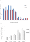
Figure 1
Figure 1
(A) MTT assays for human umbilical vein endothelial cells (HUVECs) and human mesenchymal stem cells (hMSCs) under arsenite treatment show a distinct difference in the survival rate between HUVECs and hMSCs. There are significantly higher survival rates in hMSCs than in HUVECs under high concentrations of arsenite between 15 to 25 μM; (B) An apoptosis flow cytometry study reveals significant differences in apoptosis rates between HUVECs treated with arsenite, HUVECs in conditioned medium and HUVECs and hMSCs co-cultured under an arsenite concentration of 20 μM. (* p < 0.05), MTT assay—(3-[4,5-dimethylthiazol-2-yl]-2,5-diphenyltetrazolium bromides). Col-cul—co-cultured HUVEC and hMSCs. CM—conditioned medium.
Figure 2
Figure 2
(A) Microscopic view of a culture dish with 20 µM arsenite treatment for 48 h shows obvious differences in the morphology and viability of HUVECs between cells cultured alone and those co-cultured with hMSCs. The left picture shows that when we delivered VEGF (vascular endothelia growth factor) siRNA into hMSCs, the harvested siRNA conditioned medium could not rescue the HUVECs and more cell death was found compared with the middle picture. (Scale bar 100 µm) (B) Apoptosis flow cytometry results show fewer HUVECs deviating to zone 1 and zone 4 (Annexin V positive) when cultured with conditioned media from hMSCs. This phenomenon is abolished when we deliver siRNA into hMSCs. (* p < 0.05) (C) Cytokine array assay reveals an elevated VEGF level in conditioned media with hMSCs treated with As compared with that in normal medium. (D) Western blot experiments show the same result. More VEGF was expressed upon As treatment in the conditioned medium than in the normal medium (hMSCs only). CM—conditioned medium with HUVECs treated with 20 µm As. CM+Si—conditioned medium with HUVECs and the addition of VEGF SiRNA. hMSCs—human mesenchymal stem cells. HUSMCs—human umbilical vein smooth muscle cells.
Figure 3
Figure 3
(A) A mitochondrial maximum oxygen consumption rate (OCR) test shows that with Seahorse XF-24, the OCR of hMSCs remains constant without significant changes before or after treatment with As. However, the OCR of HUVECs decreases significantly at a high concentration of As and the difference is statically significant (* p < 0.05). Cells were sequentially treated with oligomycin (line B), carbonyl cyanide p-trifluoromethoxy-phenylhydrazone (FCCP) (line C), as well as rotenone and antimycin A (line D). Maximal respiratory rate was measured after cells were treated with FCCP (blue bar zone 29). Dark orange line—HUVEC, Light orange line—HUVEC with As 20 μM. Red line—MSC, Blue line—MSC with As 20 μM. (B) A mitochondrial mass nonyl acridine orange (NAO) fluorescence assay demonstrates that under 20 µM As, the NAO green fluorescent protein (GFP) mean in HUVECs decreased compared with that in normal HUVECs. hMSCs maintain a constant mean before and after treatment with As. (C) A reactive oxygen species (ROS) assay demonstrates that ROS levels declined slightly in HUVECs treated with conditioned medium compared with those in cells treated with As for only 48 h. (D) An antioxidant superoxidase dismutase (SOD) assay shows increased SOD activity in HUVECs treated with conditioned medium compared with that in cells treated with As only. hMSCs—human mesenchymal stem cells. HUVECs—human umbilical vein endothelial cells.
Figure 3
Figure 3
(A) A mitochondrial maximum oxygen consumption rate (OCR) test shows that with Seahorse XF-24, the OCR of hMSCs remains constant without significant changes before or after treatment with As. However, the OCR of HUVECs decreases significantly at a high concentration of As and the difference is statically significant (* p < 0.05). Cells were sequentially treated with oligomycin (line B), carbonyl cyanide p-trifluoromethoxy-phenylhydrazone (FCCP) (line C), as well as rotenone and antimycin A (line D). Maximal respiratory rate was measured after cells were treated with FCCP (blue bar zone 29). Dark orange line—HUVEC, Light orange line—HUVEC with As 20 μM. Red line—MSC, Blue line—MSC with As 20 μM. (B) A mitochondrial mass nonyl acridine orange (NAO) fluorescence assay demonstrates that under 20 µM As, the NAO green fluorescent protein (GFP) mean in HUVECs decreased compared with that in normal HUVECs. hMSCs maintain a constant mean before and after treatment with As. (C) A reactive oxygen species (ROS) assay demonstrates that ROS levels declined slightly in HUVECs treated with conditioned medium compared with those in cells treated with As for only 48 h. (D) An antioxidant superoxidase dismutase (SOD) assay shows increased SOD activity in HUVECs treated with conditioned medium compared with that in cells treated with As only. hMSCs—human mesenchymal stem cells. HUVECs—human umbilical vein endothelial cells.
Figure 4
Figure 4
(A) HE (hematoxylin and eosin) staining for a small arteriole from the anterior lower limb of an SD rat shows a decrease in the lumen diameter of the arteriole (yellow arrow) and an increase in the number of inflammatory cells infiltrating the smooth muscles of vessel walls (green arrow) when compared with a wild-type untreated rat (left). Scale bar 200 μm. (B) IHC CD31 staining for vessels reveals a decrease in staining intensity in the As-treated group compared with that in the wild-type SD rat group (yellow arrow). Scale bar 200 μm. (C) A TUNEL assay shows numerous TUNEL-positive tissues induced by As in the control group. Compared with the control specimens, mMSCs-injected SD rat specimens show a decrease in TUNEL reactions, especially in regions containing anterior tibial vessels. Scale bar 100 μm. SD rat—Sprague Dawley rat. IHC—immunohistochemistry. CD31 stain—platelet endothelial cell adhesion molecular stain. TUNEL assay—terminal deoxynucleotidyl transferase dUTP nick end labeling assay. mMSCs—mouse mesenchymal stem cells.
Figure 4
Figure 4
(A) HE (hematoxylin and eosin) staining for a small arteriole from the anterior lower limb of an SD rat shows a decrease in the lumen diameter of the arteriole (yellow arrow) and an increase in the number of inflammatory cells infiltrating the smooth muscles of vessel walls (green arrow) when compared with a wild-type untreated rat (left). Scale bar 200 μm. (B) IHC CD31 staining for vessels reveals a decrease in staining intensity in the As-treated group compared with that in the wild-type SD rat group (yellow arrow). Scale bar 200 μm. (C) A TUNEL assay shows numerous TUNEL-positive tissues induced by As in the control group. Compared with the control specimens, mMSCs-injected SD rat specimens show a decrease in TUNEL reactions, especially in regions containing anterior tibial vessels. Scale bar 100 μm. SD rat—Sprague Dawley rat. IHC—immunohistochemistry. CD31 stain—platelet endothelial cell adhesion molecular stain. TUNEL assay—terminal deoxynucleotidyl transferase dUTP nick end labeling assay. mMSCs—mouse mesenchymal stem cells.
Figure 5
Figure 5
(A) An in vivo ELISA VEGF test demonstrates that VEGF levels significantly increase in blood samples drawn from SD rats with serial mMSCs (mouse MSCs) injections for two weeks (* p < 0.01). (B) An in vivo VEGF qPCR test shows increasing gene expression in rats after serial mMSCs injections for two weeks. (* p < 0.05). (C) An in vivo GSH/GSSG assay shows that the GSH/GSSG ratio increases in experimental groups subjected to serial mMSCs injections for two weeks compared with that in the As-treated group (* p < 0.05). (D) In vivo antioxidant superoxidase dismutase (SOD) assay shows increased SOD activity in experimental groups subjected to serial mMSCs injections for two weeks compared with that in the As-treated group (* p < 0.05). VEGF—vascular endothelial growth factor. mMSCs—mouse mesenchymal stem cells. PBS—phosphate buffered saline. MSC 1w—mouse MSCs injected for one week. MSC 2w—mouse MSCs injected for two weeks. GSH—reduced glutathione. GSSG—oxidized glutathione.

Similar articles

Cited by

References

    1. McCarty K.M., Hanh H.T., Kim K.W. Arsenic geochemistry and human health in South East Asia. Rev. Environ. Health. 2011;26:71–78. doi: 10.1515/reveh.2011.010. - DOI - PMC - PubMed
    1. Chung J.Y., Yu S.D., Hong Y.S. Environmental source of arsenic exposure. J. Prev. Med. Public Health. 2014;47:253–257. doi: 10.3961/jpmph.14.036. - DOI - PMC - PubMed
    1. Tseng C.H. Cardiovascular disease in arsenic-exposed subjects living in the arseniasis-hyperendemic areas in Taiwan. Atherosclerosis. 2008;199:12–18. doi: 10.1016/j.atherosclerosis.2008.02.013. - DOI - PubMed
    1. Prozialeck W.C., Edwards J.R., Nebert D.W., Woods J.M., Barchowsky A., Atchison W.D. The vascular system as a target of metal toxicity. Toxicol. Sci. Off. J. Soc. Toxicol. 2008;102:207–218. doi: 10.1093/toxsci/kfm263. - DOI - PMC - PubMed
    1. Wu M.M., Chiou H.Y., Wang T.W., Hsueh Y.M., Wang I.H., Chen C.J., Lee T.C. Association of blood arsenic levels with increased reactive oxidants and decreased antioxidant capacity in a human population of northeastern Taiwan. Environ. Health Perspect. 2001;109:1011–1017. doi: 10.1289/ehp.011091011. - DOI - PMC - PubMed

MeSH terms