

New Gene Markers for Metabolic Processes and Homeostasis in Porcine Buccal Pouch Mucosa during Cells Long Term-Cultivation-A Primary Culture Approach
- PMID: 29596348
- PMCID: PMC5979461
- DOI: 10.3390/ijms19041027
New Gene Markers for Metabolic Processes and Homeostasis in Porcine Buccal Pouch Mucosa during Cells Long Term-Cultivation-A Primary Culture Approach
Abstract
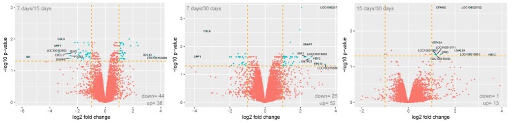
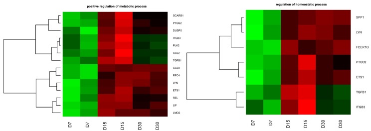
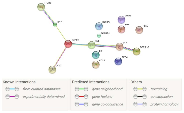
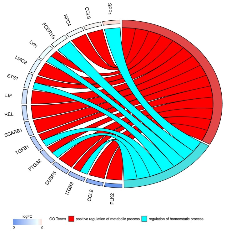
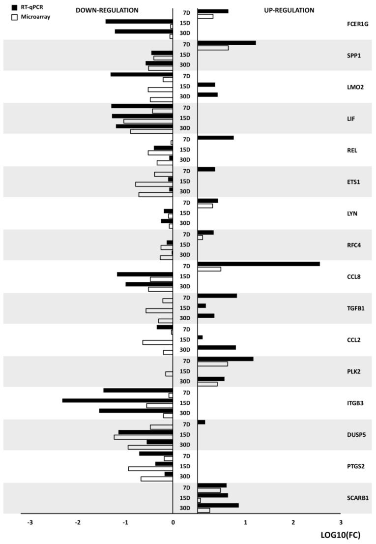
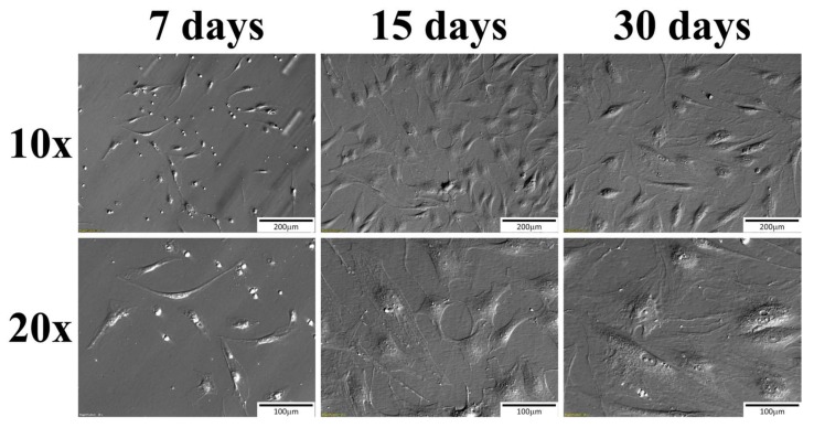
The oral mucosal tissue is a compound structure composed of morphologically and physiologically different cell types. The morphological modification involves genetically determined lifespan, which may be recognized as the balance between cell survival and apoptosis. Although the biochemical processes and pathways in oral mucosa, with special regards to drug transport, delivery, and metabolism, are well known, the cellular physiological homeostasis in this tissue requires further investigation. The porcine buccal pouch mucosal cells (BPMCs) collected from 20 pubertal crossbred Landrace gilts, were used in this study. Immediately after recovery, the oral mucosa was separated micro-surgically, and treated enzymatically. The dispersed cells were transferred into primary in vitro culture systems for a long-term cultivation of 30 days. After each step of in vitro culture (IVC), the cells were collected for isolation of total RNA at 24 h, 7, 15, and 30 days of IVC. While the expression was analyzed for days 7, 15, and 30, the 24th hour was used as a reference for outcome calibration. The gene expression profile was determined using Affymetrix microarray assays and necessary procedures. In results, we observed significant up-regulation of SCARB1, PTGS2, DUSP5, ITGB3, PLK2, CCL2, TGFB1, CCL8, RFC4, LYN, ETS1, REL, LIF, SPP1, and FGER1G genes, belonging to two ontological groups, namely "positive regulation of metabolic process", and "regulation of homeostatic process" at 7 day of IVC as compared to down-regulation at days 15 and 30. These findings suggest that the metabolic processes and homeostatic regulations are much more intense in porcine mucosal cells at day 7 of IVC. Moreover, the increased expression of marker genes, for both of these ontological groups, may suggest the existence of not only "morphological lifespan" during tissue keratinization, but also "physiological checkpoint" dedicated to metabolic processes in oral mucosa. This knowledge may be useful for preclinical experiments with drugs delivery and metabolism in both animals and humans.
Keywords: homeostasis; metabolic process; oral mucosa.
Conflict of interest statement
All authors declare they have no conflicts of interest.
Figures
Similar articles
-
Genes involved in regulation of cellular metabolic processes, signaling and adhesion are the markers of porcine buccal pouch mucosal cells long-term primary cultured in vitro.J Biol Regul Homeost Agents. 2018 Sep-Oct;32(5):1129-1141. J Biol Regul Homeost Agents. 2018. PMID: 30334405
-
Expression of genes responsible for cell morphogenesis involved in differentiation in porcine buccal pouch mucosal cells during long-term primary culture and real-time proliferation in vitro.J Biol Regul Homeost Agents. 2017 Oct-Dec;31(4):855-864. J Biol Regul Homeost Agents. 2017. PMID: 29254288
-
Differential expression and distribution of cytokeratins and vimentin in buccal pouch mucosal cells during real-time cell proliferation: research based on a porcine model.J Biol Regul Homeost Agents. 2016 Oct-Dec;30(4):951-960. J Biol Regul Homeost Agents. 2016. PMID: 28078841
-
The biomedical aspects of oral mucosal epithelial cell culture in mammals.J Biol Regul Homeost Agents. 2017 Jan-Mar;31(1):81-85. J Biol Regul Homeost Agents. 2017. PMID: 28337874 Review.
-
Cytokeratin changes in cell culture systems of epithelial cells isolated from oral mucosa: a short review.Ital J Anat Embryol. 2005 Apr-Jun;110(2):75-82. Ital J Anat Embryol. 2005. PMID: 16277157 Review.
Cited by
-
Transcriptomic analysis of expression of genes regulating cell cycle progression in porcine ovarian granulosa cells during short-term in vitro primary culture.Histochem Cell Biol. 2020 Jun;153(6):397-412. doi: 10.1007/s00418-020-01860-2. Epub 2020 Mar 10. Histochem Cell Biol. 2020. PMID: 32157392 Free PMC article.
-
Expression Profile of New Marker Genes Involved in Differentiation of Canine Adipose-Derived Stem Cells into Osteoblasts.Int J Mol Sci. 2021 Jun 22;22(13):6663. doi: 10.3390/ijms22136663. Int J Mol Sci. 2021. PMID: 34206369 Free PMC article.
-
Expression Profile of New Gene Markers Involved in Differentiation of Canine Adipose-Derived Stem Cells into Chondrocytes.Genes (Basel). 2022 Sep 16;13(9):1664. doi: 10.3390/genes13091664. Genes (Basel). 2022. PMID: 36140831 Free PMC article.
References
-
- Borys S., Khozmi R., Kranc W., Bryja A., Dyszkiewicz-Konwińska M., Jeseta M., Kempisty B. Recent findings of the types of programmed cell death. Adv. Cell Biol. 2017;5:43–49. doi: 10.1515/acb-2017-0004. - DOI
-
- Bryja A., Dyszkiewicz-Konwińska M., Budna J., Ciesiółka S., Kranc W., Borys S., Jeseta M., Urbaniak O., Bukowska D., Antosik P., et al. Expression of cell mitotic progression proteins and keratinocyte markers in porcine buccal pouch mucosal cells during short-term, real-time primary culture. J. Biol. Regul. Homeost. Agents. 2017;31:297–309. - PubMed
-
- Bryja A., Dyszkiewicz-Konwińska M., Budna J., Kranc W., Chachuła A., Borys S., Ciesiółka S., Ciesiółka J., Prylinski M., Prylinski D., et al. The biomedical aspects of oral mucosal epithelial cell culture in mammals. J. Biol. Regul. Homeost. Agents. 2017;31:81–85. - PubMed
-
- Bryja A., Dyszkiewicz-Konwińska M., Chachuła A., Ciesiółka S., Kranc W., Bukowska D., Antosik P., Bruska M., Nowicki M., Zabel M., et al. Differential expression and distribution of cytokeratins and vimentin in buccal pouch mucosal cells during real-time cell proliferation: Research based on a porcine model. J. Biol. Regul. Homeost. Agents. 2016;30:951–960. - PubMed
-
- Kranc W., Celichowski P., Budna J., Khozmi R., Bryja A., Ciesiółka S., Rybska M., Borys S., Jeseta M., Bukowska D., et al. Positive Regulation of Macromolecule Metabolic Process Belongs to the Main Mechanisms Crucial for Porcine Ooocytes Maturation. Adv. Cell Biol. 2017;5:15–31. doi: 10.1515/acb-2017-0002. - DOI
MeSH terms
LinkOut - more resources
Full Text Sources
Other Literature Sources
Research Materials
Miscellaneous
