. 2018 Mar 29;11(4):521.
doi: 10.3390/ma11040521.

Porous Polyethylene Coated with Functionalized Hydroxyapatite Particles as a Bone Reconstruction Material

Affiliations

Porous Polyethylene Coated with Functionalized Hydroxyapatite Particles as a Bone Reconstruction Material

H Fouad et al. Materials (Basel). .

Abstract

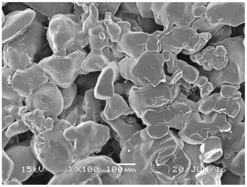
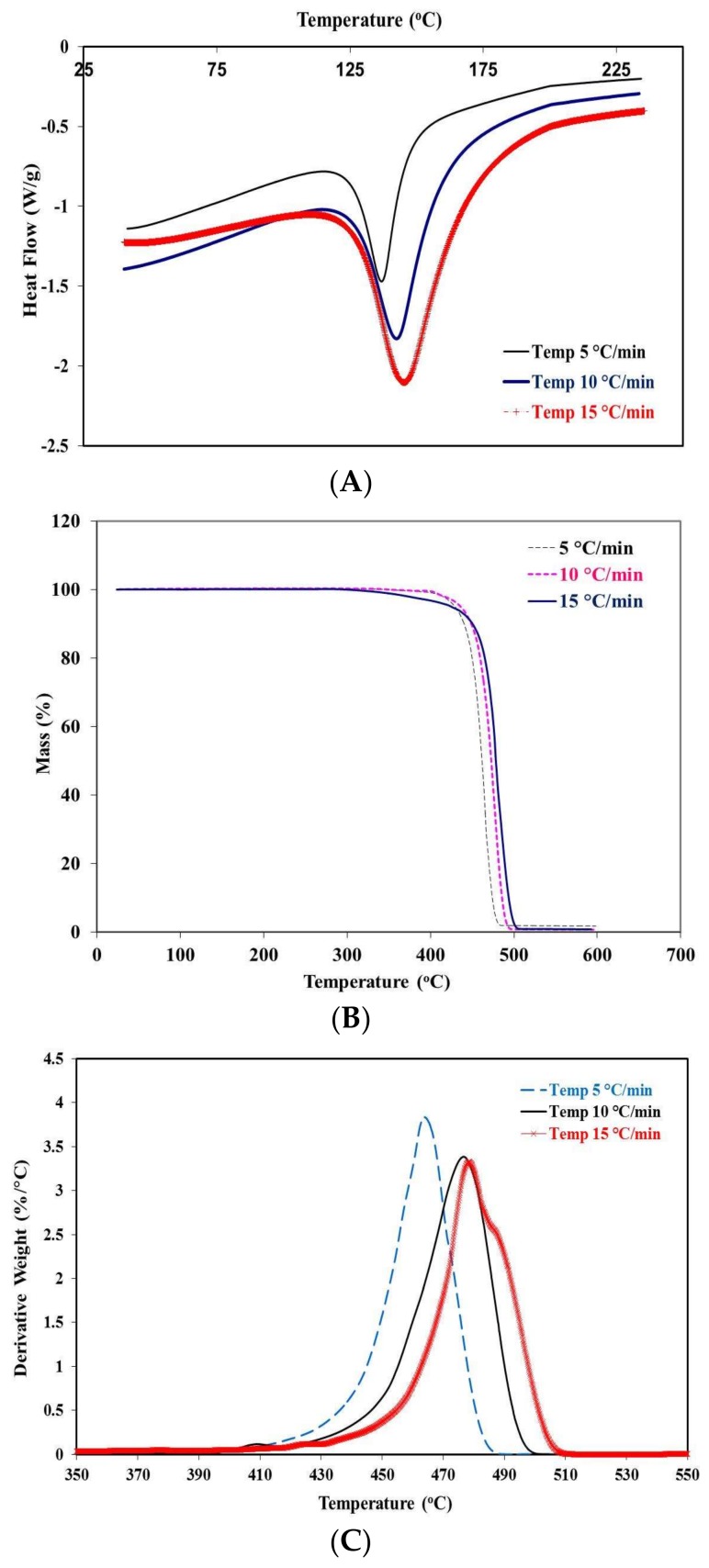
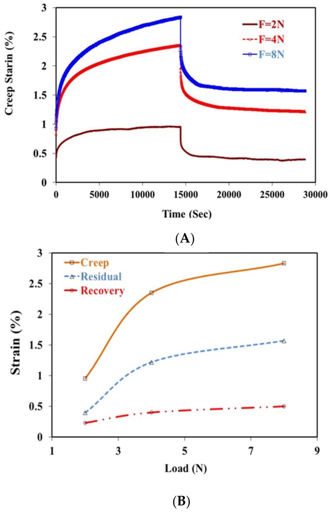
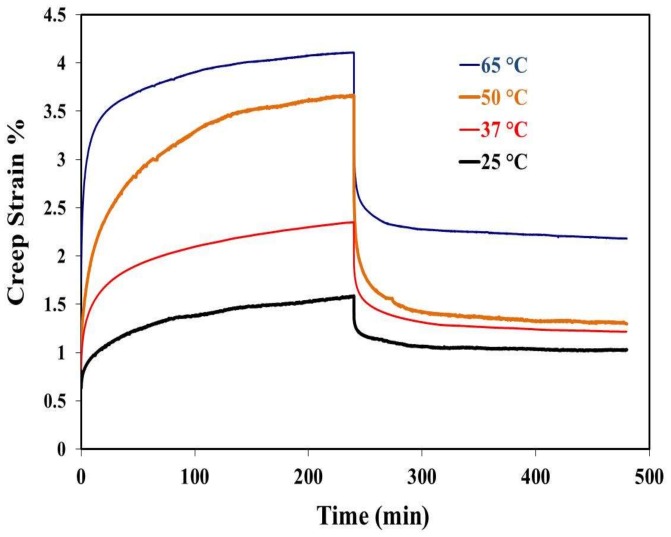
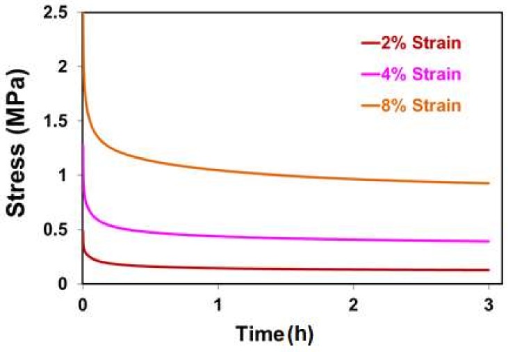
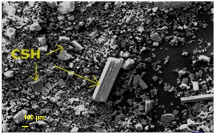
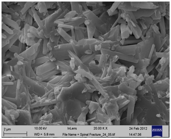
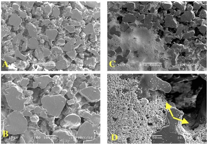
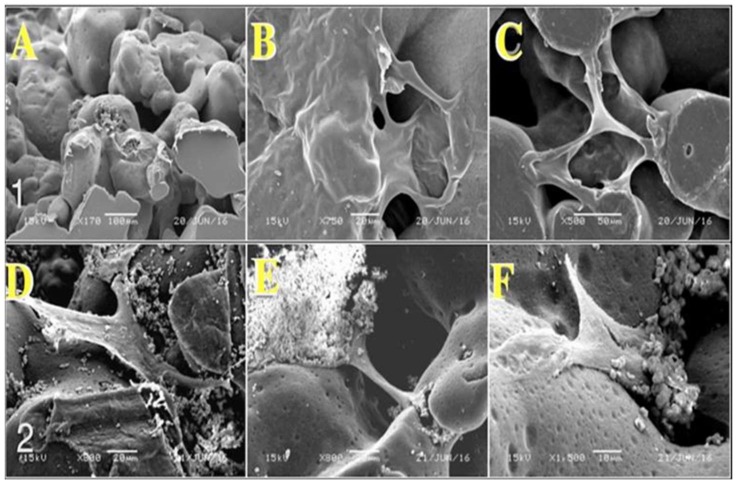
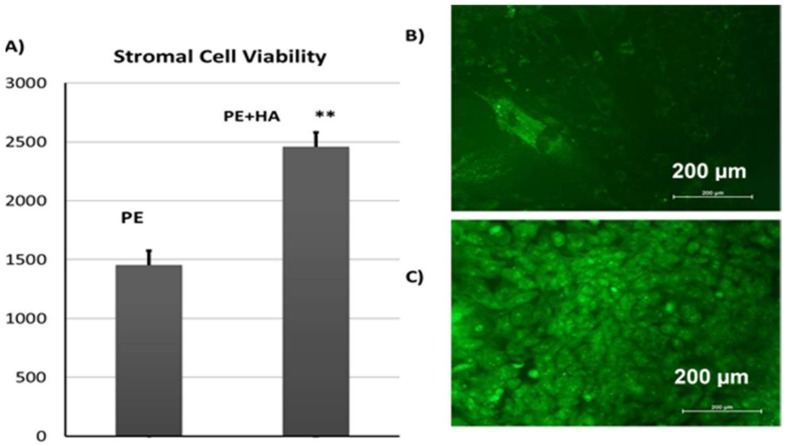
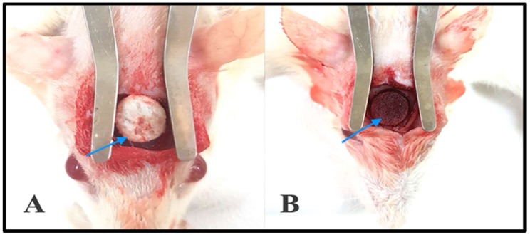
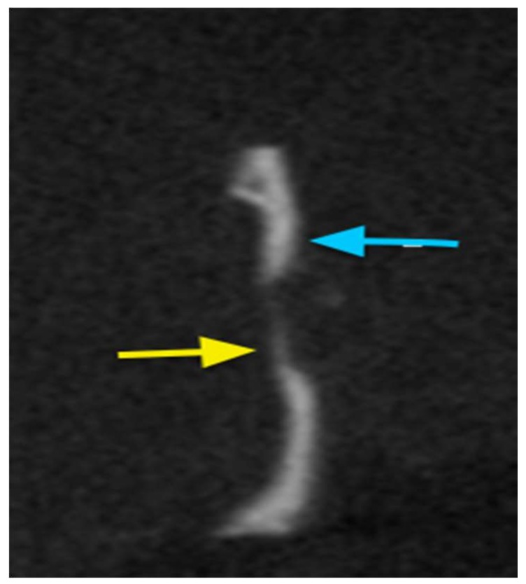
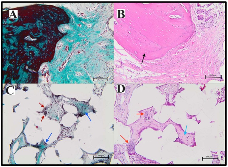
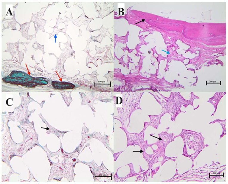
In this study, porous polyethylene scaffolds were examined as bone substitutes in vitro and in vivo in critical-sized calvarial bone defects in transgenic Sprague-Dawley rats. A microscopic examination revealed that the pores appeared to be interconnected across the material, making them suitable for cell growth. The creep recovery behavior of porous polyethylene at different loads indicated that the creep strain had two main portions. In both portions, strain increased with increased applied load and temperature. In terms of the thermographic behavior of the material, remarkable changes in melting temperature and heat fusion were revealed with increased the heating rates. The tensile strength results showed that the material was sensitive to the strain rate and that there was adequate mechanical strength to support cell growth. The in vitro cell culture results showed that human bone marrow mesenchymal stem cells attached to the porous polyethylene scaffold. Calcium sulfate-hydroxyapatite (CS-HA) coating of the scaffold not only improved attachment but also increased the proliferation of human bone marrow mesenchymal stem cells. In vivo, histological analysis showed that the study groups had active bone remodeling at the border of the defect. Bone regeneration at the border was also evident, which confirmed that the polyethylene acted as an osteoconductive bone graft. Furthermore, bone formation inside the pores of the coated polyethylene was also noted, which would enhance the process of osteointegration.

Keywords: hydroxyapatite; mesenchymal stem cells; porous polyethylene.


Conflict of interest statement

The authors declare that there are no conflicts of interest or state among all the contributors.

Figures

Figure 1
Figure 1
Scanning electron microscopic images of the porous polyethylene. Surface scale bar = 100 μm.
Figure 2
Figure 2
(A) Differential scanning calorimetry (DSC); (B,C) thermogravimetric analysis (TGA) results for porous polyethylene (PE) at different heating rates.
Figure 3
Figure 3
Effects of load on the creep behavior of porous polyethylene.
Figure 4
Figure 4
Effects of temperature on the creep behavior of porous polyethylene.
Figure 5
Figure 5
Relaxation behavior of porous polyethylene at different initial strains.
Figure 6
Figure 6
Microradiograph for scanning electron microscopy of calcium sulfate hydrate–hydroxyapatite powder showing calcium sulfate hydrate in rectangular and rod-like shapes (arrows) and smaller particles of hydroxyapatite. Scale bar = 100 μm. calcium sulfate hydrate.
Figure 7
Figure 7
Microradiograph for scanning electron microscopy of calcium sulfate hydrate–hydroxyapatite, which attained compact structures of well-interconnected crystals. The hydroxyapatite crystals are shorter than the calcium sulfate hydrate crystals. Scale bar = 2 μm.
Figure 8
Figure 8
Microradiograph for scanning electron microscopy of porous polyethylene. (A,B) show the cut section of non-coated porous polyethylene; the variation in pore size is noted to vary within the range of 50–400 μm. (A,B) scale bars = 500 and 100 μm. (C,D) show the coated porous polyethylene.
Figure 9
Figure 9
Micrograph of porous polyethylene three days after cell seeding. The first row represents the cell culture on uncoated porous polyethylene discs; cells are mainly found toward the edge of the discs (A). Cells are also found crossing the scaffold’s pores (AC). Scale bars = 100, 20, and 50 μm. (DF) show cells with the typical morphology of stromal cells (polygonal or fusiform). The cells are intermingled with coating material’s crystals, and crystalline apatite on the surface of material can be seen. Cells migrated inside the pores of porous polyethylene. Scale bars = 20, 20, and 10 μm.
Figure 10
Figure 10
(A) AlamarBlue® assay of CL1 cells cultured on porous polyethylene and polyethylene + hydroxylapatite scaffolds; (B) CL1 cells grown on a polyethylene scaffold; (C) CL2 cells grown on polyethylene + hydroxylapatite scaffolds stained with acridine orange, which stains all nucleated cells green. The magnification used was 10×.
Figure 11
Figure 11
Photographs showing the surgical defects at the rat calvarium. (A) Shows the coated porous polyethylene disc in situ; (B) Shows the untreated polyethylene disc in situ.
Figure 12
Figure 12
Sagittal view for cone beam computed tomography (CBCT) for the coated PE disc in situ after three months. The area of bone regeneration is shown by the yellow arrow, while the area of native bone is shown by the blue arrow. The area of the PE graft appears radiolucent.
Figure 13
Figure 13
Photomicrographs of decalcified sections stained with hematoxylin and eosin (right) and Masson’s trichrome stain (left) demonstrating the area of bone regeneration at the surgical defect using coated porous polyethylene. (A,B) are sections through the area at the interface between the bone border of the defect and the implanted coated polyethylene. The bone shows active remodeling, and the connective tissue next to the bone is highly rich with collagen (green) and osteoblasts and preosteoblast-like cells. (C,D) show sections from the center of the scaffold; the interpore spaces are full of connective tissue that turned into bone and osteoid (C,D blue arrows). The presence of hydroxylapatite is noted (D, red arrow). There are also giant cells seen mainly in this section (C, red arrow).
Figure 14
Figure 14
H&E and Masson’s trichrome staining for non-coated polyethylene disks. Decalcified sections were stained with hematoxylin and eosin (right) and Masson’s trichrome stain (left), demonstrating the area of bone regeneration at the surgical defect using plain polyethylene. (A,B) are sections through the area of the peripheral bone defect and the implanted plain non-coated polyethylene. (B) Black arrow shows newly generated bone with evidence of active remodeling. The connective tissue is next to the bone, with osteoblastic and pre-osteoblast like cells (see blue arrows). (C,D) are sections from the center of the scaffold; the interpore spaces are full of connective tissue that turned into bone in the osteons (C,D black arrow).

Similar articles

Cited by

References

    1. Jeremy M.H., Peter X.M. Biomimetic nanofibrous scaffolds for bone tissue engineering. Biomaterials. 2011;32:9622–9629. - PMC - PubMed
    1. Behnaz B., Payam Z., Mohammad O.O., Farid K., Hamideh F., Sohrabi-Jahromi S., Zarrintaj Z. Tissue engineering; strategies, tissues, and biomaterials. Biotechnol. Genet. Eng. Rev. 2017;33:144–172. - PubMed
    1. Schlickewei W., Schlickewei S. The use of bone substitutes in the treatment of bone defects—The clinical view and history. Macromol. Symp. 2007;253:10–23. doi: 10.1002/masy.200750702. - DOI
    1. Stevens M.M. Biomaterials for bone tissue engineering. Mater. Today. 2008;11:18–25. doi: 10.1016/S1369-7021(08)70086-5. - DOI
    1. Chen C.T., Hu T.L., Lai J.B., Chen Y.C., Chen Y.R. Reconstruction of traumatic nasal deformity in Orientals. J. Plast. Reconstr. Aesthet. Surg. 2010;63:257–264. doi: 10.1016/j.bjps.2008.10.006. - DOI - PubMed