. 2018 Mar 29;16(4):111.
doi: 10.3390/md16040111.

Production, Characterization and Biocompatibility Evaluation of Collagen Membranes Derived from Marine Sponge Chondrosia reniformis Nardo, 1847

Affiliations

Production, Characterization and Biocompatibility Evaluation of Collagen Membranes Derived from Marine Sponge Chondrosia reniformis Nardo, 1847

Marina Pozzolini et al. Mar Drugs. .

Abstract

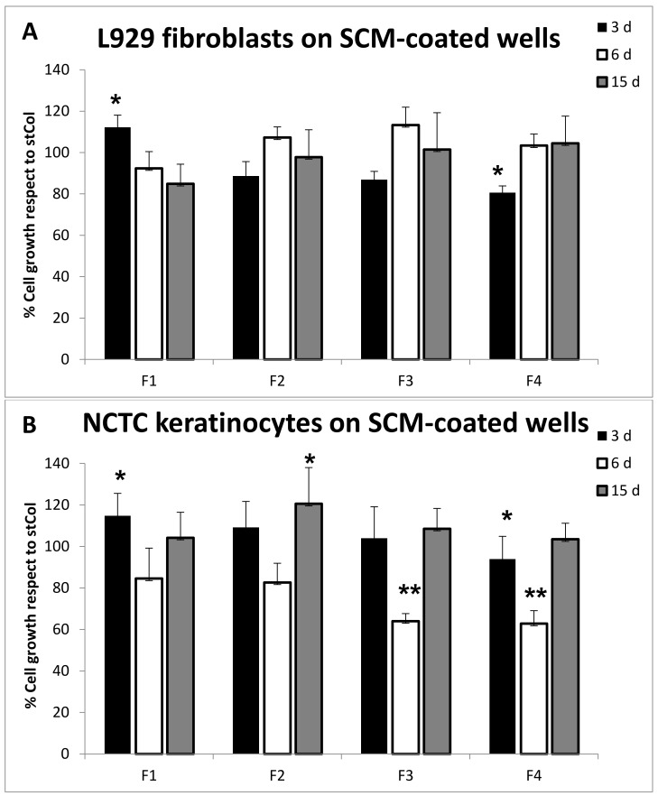
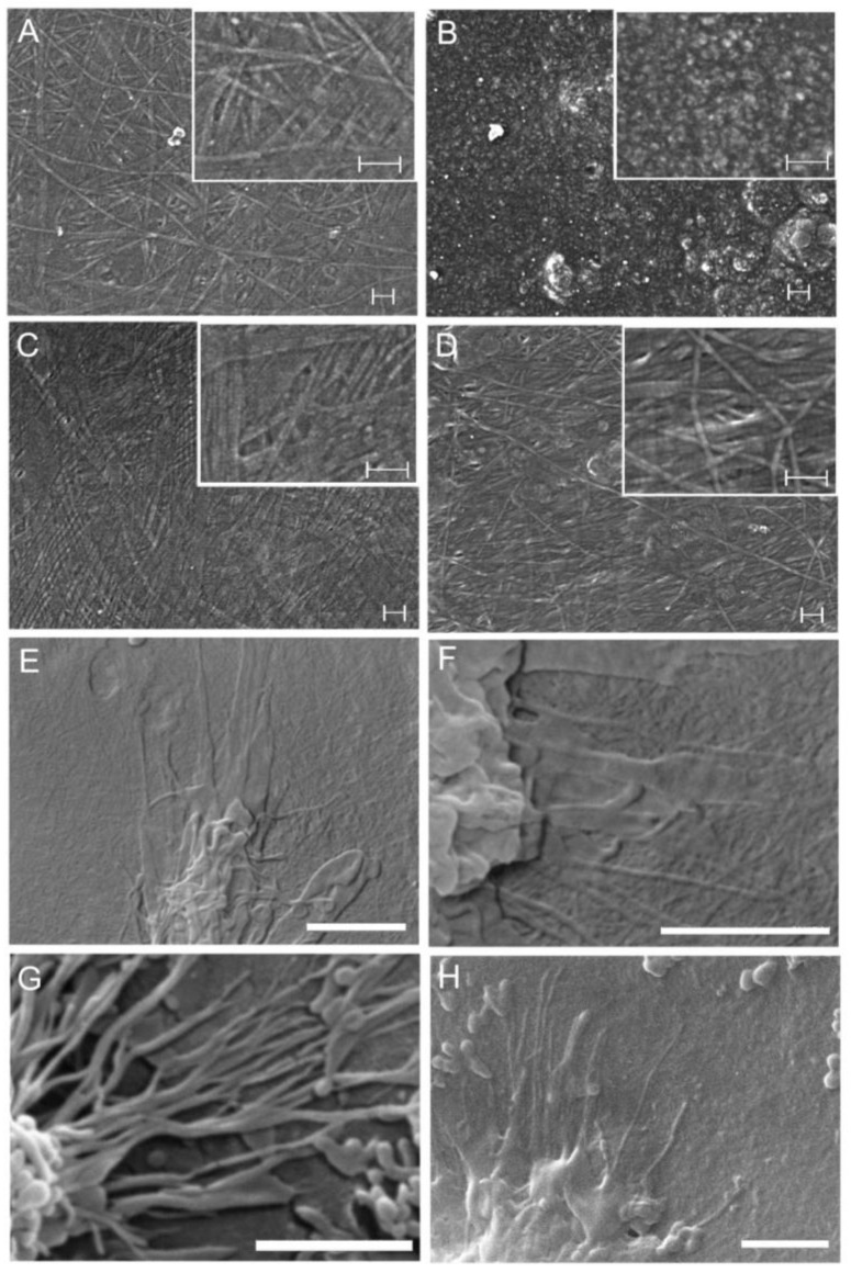
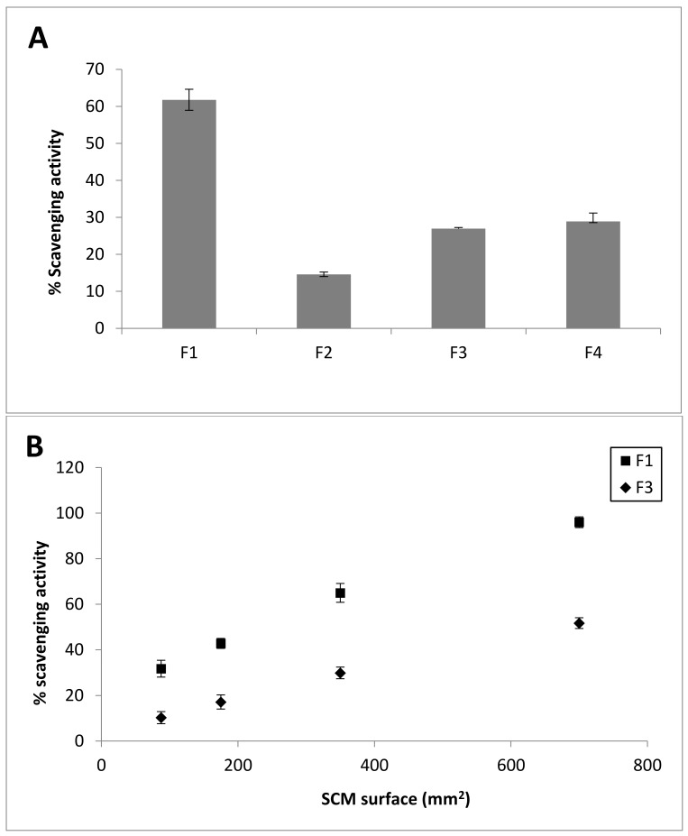
Collagen is involved in the formation of complex fibrillar networks, providing the structural integrity of tissues. Its low immunogenicity and mechanical properties make this molecule a biomaterial that is extremely suitable for tissue engineering and regenerative medicine (TERM) strategies in human health issues. Here, for the first time, we performed a thorough screening of four different methods to obtain sponge collagenous fibrillar suspensions (FSs) from C. reniformis demosponge, which were then chemically, physically, and biologically characterized, in terms of protein, collagen, and glycosaminoglycans content, viscous properties, biocompatibility, and antioxidant activity. These four FSs were then tested for their capability to generate crosslinked or not thin sponge collagenous membranes (SCMs) that are suitable for TERM purposes. Two types of FSs, of the four tested, were able to generate SCMs, either from crosslinking or not, and showed good mechanical properties, enzymatic degradation resistance, water binding capacity, antioxidant activity, and biocompatibility on both fibroblast and keratinocyte cell cultures. Finally, our results demonstrate that it is possible to adapt the extraction procedure in order to alternatively improve the mechanical properties or the antioxidant performances of the derived biomaterial, depending on the application requirements, thanks to the versatility of C. reniformis extracellular matrix extracts.

Keywords: Porifera; biomaterials; collagen; membranes; tissue engineering.


Conflict of interest statement

The authors declare no conflict of interest.

Figures

Scheme 1
Scheme 1
Extraction methods used. Schematic representation of the four extraction procedures used to obtain four different sponge fibrillar suspensions (F1–F4) as detailed in Section 4.1. Method 1 from [53]; method 2 from [54]; method 3 from [46]; and, method 4 from [55].
Figure 1
Figure 1
Fibrillar suspensions (FS) appearance and viscosity test. (A) Appearance of F1-F4 sponge FSs obtained through the four different extraction methods summarized in Scheme 1; (B) Flow sweep curves obtained in the viscosity tests of the four FSs by rheological measurements (for methods see Section 4.3.6). Curves were fitted by the Carreau-Gahleitner model (Mezger, 2006) as shown in Equation: ηηη0η= 1(1+(a· γ˙)b)P where η is the shear viscosity, η is the infinity-shear viscosity, η0 is the zero-shear viscosity, a is the Carreau constant, b is the Gahleitner exponent, and P is the Carreau exponent. The experimental values of η0 and η are shown in Table 3.
Figure 2
Figure 2
TEM analysis. Representative negatively stained transmission electron microscopy images of the four FSs (F1–F4) taken at 92,000× magnification (scale bar: 200 nm) with a CM10 Philips transmission electron microscope equipped with Megaview 3 camera and Olympus SIS iTEM software for digital image acquisition. (A) F1; (B) F2; (C) F3; and, (D) F4. All of the FSs show long unbranched banded fibrils of uniform size and periodicity band pattern. F1, F2, and F4 show small clots and filaments (white arrows) of putative mucopolysaccharides.
Figure 3
Figure 3
FS histological staining. The four FSs (F1–F4) smeared on histological slides and stained according to four different staining procedures, as described in Section 4.3.4, and observed by optical/polarized microscopy. (AD) Alcian pH 2.5 staining, highlighting mucopolysaccharides (MPS) and glycoproteins in blue, by reacting with acidic groups; (EH) Periodic Acid Schiff (Hotchkiss–Mc Manus) (PAS)_staining highlighting MPS, glycoproteins, glycolipids, mucins, and polysaccharides such as glycogen in pink/red, by reacting with basic groups; (IL) Picro-Sirius Red staining of collagen bundles in different shades of green, red, or yellow, depending on the thickness and the packing of fibres, from thinner to thicker, respectively. Each column of panels represents the various histological stainings of each FS. Scale bars in each panel span 20 micrometer.
Figure 4
Figure 4
Sponge Collagen Membranes (SCMs). (A) SCMs derived from F1–F4 sponge extracts crosslinked with 1-ethyl-3-(3-dimethylaminopropyl) carbodiimide/N-hydroxysuccinimide (EDC/NHS) solution; (B) not crosslinked SCMs derived from F1 and F3 sponge extracts. Scale bars in each panel span 2.0 cm.
Figure 5
Figure 5
The elastic moduli G′ (filled symbols) and the viscous moduli G′′ (open symbols) of the SCMs (SCM-F1, triangles; SCM-F3, stars) and not crosslinked SCMs (nc-SCM-F1, circles; nc-SCM-F3, diamonds) reported in function of the frequency applied by the rheometer in the dynamic mechanical analysis (for methods see Section 4.5.3).
Figure 6
Figure 6
SCM serum interaction and water absorbing capacity. (A) SCM-F1 incubated in 1 mL of FBS (left) or PBS (right) for 15 day at 37 °C; (B) Stereo-microscope observation of SCM-F1after incubation for 15 days at 37 °C in FBS (left) or PBS (right); scale bar, 50 micrometer; (C) Not cross-linked (nc-SCM) and cross-linked SCM derived from F1 and F3 fibrillar extracts after soaking in PBS for 1 h.
Figure 7
Figure 7
Cell adhesion evaluation. (A) Cell adhesion qualitative evaluation, by optical microscopy, of crystal violet stained L929 fibroblasts (first row) and National Collection of Type Cultures (NCTC) keratinocytes (second row) on the four different FS pre-coated plates (F1–F4) after 16 h incubation. Scale bars, 20 micrometer; (B) Cell adhesion quantitative evaluation, by MTT test, of L929 fibroblasts (black bars) and NCTC keratinocytes (grey bars) on the four different FS pre-coated plates after 16 h incubation. Results are expressed as cell percentages with respect to controls that were seeded on standard rat tail collagen-coated (stCol) wells and are the mean ± S.D. of two experiments that were performed in quadruplicate. Asterisks indicate significance in Tukey test (black bars ANOVA, p < 0.05; Tukey, F4 vs. stCol p < 0.05).
Figure 8
Figure 8
Cell growth evaluation. (A) L929 fibroblast cell growth evaluation, by MTT test, on the four different FS pre-coated plates (F1–F4) after 3 day (black bars), 6 day (white bars) and 15 day (grey bars) incubation. Results are expressed as cell percentages respect to controls seeded on standard rat tail collagen-coated (stCol) wells and are the mean ± S.D. of two experiments performed in quadruplicate. Asterisks indicate significance in Tukey test (black bars ANOVA, p < 0.0001; white bars ANOVA, p < 0.0001; grey bars ANOVA, p < 0.05; Tukey vs. stCol: * p < 0.05, ** p < 0.001, respectively); (B) NCTC keratinocytes cell growth evaluation, by MTT test, on the four different FS pre-coated plates after 3 day (black bars), 6 day (white bars), and 15 day (grey bars) incubation. Results are expressed as cell percentages with respect to controls that are seeded on standard rat tail collagen-coated (stCol) wells and are the mean ± S.D. of two experiments that were performed in quadruplicate. Asterisks indicate significance in Tukey test (black bars ANOVA, p < 0.00001; Tukey vs. stCol: * p < 0.05).
Figure 9
Figure 9
Environmental Scanning Electron Microscope (ESEM) analysis. All crosslinked SCMs (AD), dehydrated at critical point and graphite covered, were observed with a FESEM Zeiss SUPRA 40 VP instrument, while only SCM-F1 and SCM-F3, in the presence of fibroblasts and keratinocytes (EH) dehydrated at critical point and graphite covered, were observed with an ESEM Vega3–Tescan instrument. (A) SCM-F1; (B) SCM-F2; (C) SCM-F3; (D) SCM-F4; (E,F) Visualization of L929 fibroblasts adhesion to the two SCMs; (E) SCM-F1; (F) SCM-F3; (G,H) Visualization of NCTC keratinocytes adhesion to the two SCMs; (G) SCM-F1; (H) SCM-F3. In (AD) scale bars span 200 nm; in (EH) scale bars span 2 micrometer.
Figure 10
Figure 10
FS and SCM radical scavenging activity. (A) Antioxidant activity of the four different FSs (F1–F4) measured by the spectrophotometric DPPH oxidation assay using a concentration of 1 mg/mL of each FS. Results are expressed as percentages of radical scavenging activity based on the inhibition of DPPH oxidation calculated, as described in Section 4.7, and are the mean ± S.D. of two experiments performed in triplicate (ANOVA, p < 0.000001); (B) Antioxidant activity of SCM-F1 and SCM-F3 measured by the DPPH assay. Results are expressed as percentages of radical scavenging activity in function of the SCM surface area of the two membranes, and are the mean ± S.D. of two experiments performed in triplicate (ANOVA, p < 0.000001).

Similar articles

Cited by

References

    1. Ellis D.L., Yannas I.V. Recent advances in tissue synthesis in vivo by use of collagen glycosaminoglycan copolymers. Biomaterials. 1996;17:291–299. doi: 10.1016/0142-9612(96)85567-0. - DOI - PubMed
    1. Chen P., Marsilio E., Goldstein R.H., Yannas I.V., Spector M. Formation of lung alveolar like structures in collagen-glycosaminoglycan scaffolds in vitro. Tissue Eng. 2005;11:1436–1448. doi: 10.1089/ten.2005.11.1436. - DOI - PubMed
    1. Buijtenhuijs P., Buttafoco L., Poot A.A., Daamen W.F., van Kuppevelt T.H., Dijkstra P., de Vos R.A., Ster L.M., Geelkerken B.R., Feijen J., et al. Tissue engineering of bloodvessels: Characterization of smooth-muscle cells for culturing on collagen-and-elastin-based scaffolds. Biotechnol. Appl. Biochem. 2004;39:141–149. doi: 10.1042/BA20030105. - DOI - PubMed
    1. Garcia Y., Hemantkumar N., Collighan R., Griffin M., Rodriguez-Cabello J.C., Pandit A. In vitro characterization of a collagen scaffold enzymatically cross-linked with a tailored elastin-like polymer. Tissue Eng. Part A. 2009;15:887–899. doi: 10.1089/ten.tea.2008.0104. - DOI - PubMed
    1. Damour O., Gueugniaud P.Y., Berthin-Maghit M., Rousselle P., Berthod F., Sahuc F., Collombel C. A dermal substrate made of collagen-GAG-chitosan for deep burn coverage: First clinical uses. Clin. Mater. 1994;15:273–276. doi: 10.1016/0267-6605(94)90057-4. - DOI - PubMed

LinkOut - more resources