

Critical in vivo roles of WNT10A in wound healing by regulating collagen expression/synthesis in WNT10A-deficient mice
- PMID: 29596490
- PMCID: PMC5875851
- DOI: 10.1371/journal.pone.0195156
Critical in vivo roles of WNT10A in wound healing by regulating collagen expression/synthesis in WNT10A-deficient mice
Abstract
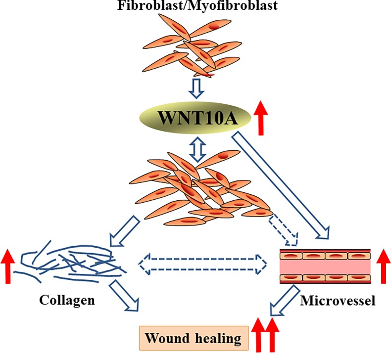
Background: We have reported that WNT10A plays a critical role in the growth of fibroblasts/myofibroblasts and microvascular endothelial cells, i.e.; wound healing/scarring. To ascertain the in vivo regulatory, central functions of WNT10A, we examined the net effects of WNT10A depletion using WNT10A-deficient mice (WNT10A-/-).
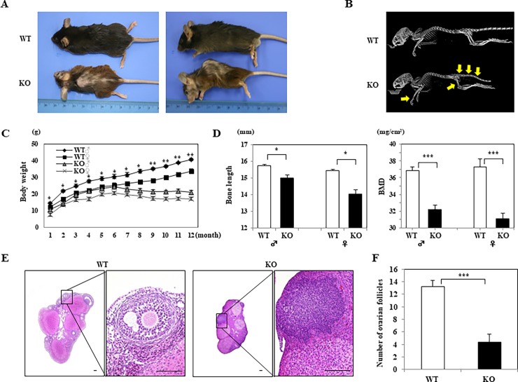
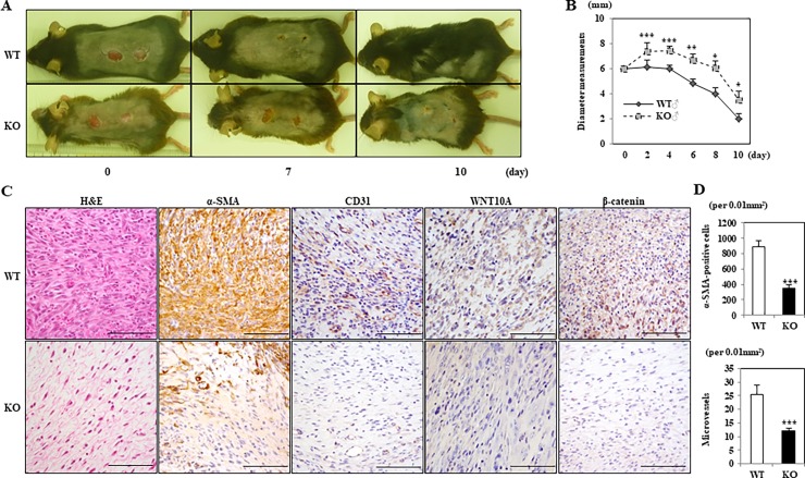
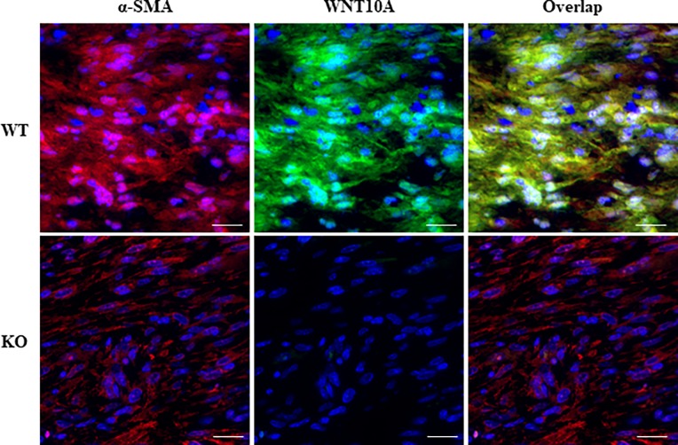
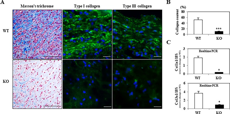
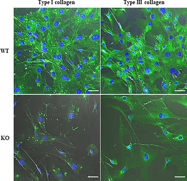
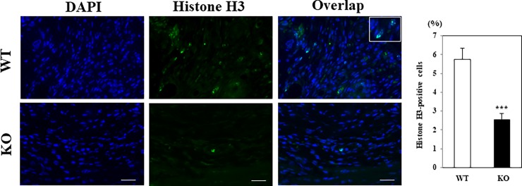
Methods and results: We generated WNT10A-/-mice, displaying a range of unique phenotypes of morpho/organogenetic failure, such as growth retardation, alopecia, kyphosis and infertility, and then focused on the functions of WNT10A in wound healing. We subjected C57BL/6J wild-type (WT) or WNT10A-/-mice to skin ulcer formation. The WNT10A-/-mice had significantly larger injured areas and delayed wound healing, which were associated with (a) a smaller number of fibroblasts/myofibroblasts and microvessels; and (b) more reduced expression and synthesis of collagen, compared with WT mice with intact WNT10A expression, especially in those with activated myofibroblasts.
Conclusions: These observations indicate that WNT10A signaling can play a pivotal in vivo role in wound healing by regulating the expression and synthesis of collagen, as one of fibrogenic factors, at least in part, and critical in vivo roles of WNT10A-mediated effective wound healing are extremely closely associated with collagen expression.
Conflict of interest statement
Figures
Similar articles
-
Depletion of WNT10A Prevents Tumor Growth by Suppressing Microvessels and Collagen Expression.Int J Med Sci. 2019 Jan 29;16(3):416-423. doi: 10.7150/ijms.26997. eCollection 2019. Int J Med Sci. 2019. PMID: 30911276 Free PMC article.
-
Neuronal PAS Domain 2 (Npas2)-Deficient Fibroblasts Accelerate Skin Wound Healing and Dermal Collagen Reconstruction.Anat Rec (Hoboken). 2020 Jun;303(6):1630-1641. doi: 10.1002/ar.24109. Epub 2019 Mar 27. Anat Rec (Hoboken). 2020. PMID: 30851151 Free PMC article.
-
Calpain activity is essential in skin wound healing and contributes to scar formation.PLoS One. 2012;7(5):e37084. doi: 10.1371/journal.pone.0037084. Epub 2012 May 16. PLoS One. 2012. PMID: 22615899 Free PMC article.
-
Role of discoidin domain receptor 2 in wound healing.Histol Histopathol. 2014 Nov;29(11):1355-64. doi: 10.14670/HH-29.1355. Epub 2014 Apr 29. Histol Histopathol. 2014. PMID: 24781958 Review.
-
Molecular Mechanisms and Potential Therapeutic Targets in Incisional Hernia.J Surg Res. 2019 Apr;236:134-143. doi: 10.1016/j.jss.2018.11.037. Epub 2018 Dec 14. J Surg Res. 2019. PMID: 30694748 Free PMC article. Review.
Cited by
-
A multiscale hybrid mathematical model of epidermal-dermal interactions during skin wound healing.Exp Dermatol. 2019 Apr;28(4):493-502. doi: 10.1111/exd.13909. Exp Dermatol. 2019. PMID: 30801791 Free PMC article.
-
Depletion of WNT10A Prevents Tumor Growth by Suppressing Microvessels and Collagen Expression.Int J Med Sci. 2019 Jan 29;16(3):416-423. doi: 10.7150/ijms.26997. eCollection 2019. Int J Med Sci. 2019. PMID: 30911276 Free PMC article.
-
Transcriptomic and Immunohistochemical Analysis of Progressive Keratoconus Reveal Altered WNT10A in Epithelium and Bowman's Layer.Invest Ophthalmol Vis Sci. 2021 May 3;62(6):16. doi: 10.1167/iovs.62.6.16. Invest Ophthalmol Vis Sci. 2021. PMID: 33988693 Free PMC article.
-
Deletion of Wnt10a Is Implicated in Hippocampal Neurodegeneration in Mice.Biomedicines. 2022 Jun 25;10(7):1500. doi: 10.3390/biomedicines10071500. Biomedicines. 2022. PMID: 35884806 Free PMC article.
-
[Research advances on the mechanism of Wnt/β-catenin signaling pathway in body surface wound healing].Zhonghua Shao Shang Yu Chuang Mian Xiu Fu Za Zhi. 2023 Feb 20;39(2):190-195. doi: 10.3760/cma.j.cn501225-20220816-00348. Zhonghua Shao Shang Yu Chuang Mian Xiu Fu Za Zhi. 2023. PMID: 36878529 Free PMC article. Chinese.
References
-
- Heise RL, Stober V, Cheluvaraju C, Hollingsworth JW, Garantziotis S. Mechanical stretch induces epithelial-mesenchymal transition in alveolar epithelia via hyaluronan activation of innate immunity. J Biol Chem. 2011;286: 17435–44. doi: 10.1074/jbc.M110.137273 - DOI - PMC - PubMed
-
- Macheda ML, Stacker SA. Importance of Wnt signaling in the tumor stroma microenvironment. Curr Cancer Drug Targets. 2008;8: 454–65. - PubMed
-
- Dvorak HF. Tumors wounds that do not heal. Similarities between tumor stroma generation and wound healing. N Engl J Med. 1986,315: 1650–9. doi: 10.1056/NEJM198612253152606 - DOI - PubMed
-
- Schäfer M, Werner S. Cancer as an overhealing wound: an old hypothesis revisited. Nat Rev Mol Cell Biol. 2008;9: 628–38. doi: 10.1038/nrm2455 - DOI - PubMed
Publication types
MeSH terms
Substances
Grants and funding
LinkOut - more resources
Full Text Sources
Other Literature Sources
Molecular Biology Databases
