

doi: 10.1371/journal.ppat.1006893.
eCollection 2018 Mar.
Hostile intruder: Toxoplasma holds host organelles captive
Affiliations
- PMID: 29596535
- PMCID: PMC5875880
- DOI: 10.1371/journal.ppat.1006893
Item in Clipboard
Hostile intruder: Toxoplasma holds host organelles captive
PLoS Pathog.
.
Erratum in
-
Correction: Hostile intruder: Toxoplasma holds host organelles captive.PLoS Pathog. 2018 Apr 24;14(4):e1007018. doi: 10.1371/journal.ppat.1007018. eCollection 2018 Apr. PLoS Pathog. 2018. PMID: 29689101 Free PMC article.
No abstract available
Conflict of interest statement
The authors have declared that no competing interests exist.
Figures
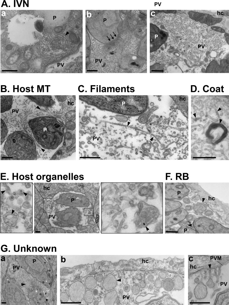
(A-G) Transmission EM of intracellular Toxoplasma. (A) Thin tubules formed by the parasite (P) 20 min post-invasion and packaged within a vesicle (panel a; arrowhead) before being discharged at the basal end of the parasite (panel b; arrows) and spread within the vacuole to form the IVN. (B) Host MT–based invaginations of the PV membrane (arrowheads). (C) Long microfilaments narrower than the IVN tubules (arrowheads). (D) IVN tubules and microtubular structures are coated by e-dense material. (E) Host endocytic organelles containing LDL-gold particles (arrowheads) surrounded by the PV membrane. (F) RB of the mother cells identified by discarded organelles such as the ER, either still attached to daughter cells or free in the PV lumen (arrowheads). (G) Unknown membrane-bound structures containing fibrillary material (arrowheads) accumulated between parasites (panel a), close to the PV membrane (panel b), or appending to the PV membrane (panel c). All scale bars, 500 nm. e-dense, electron-dense; EM, electron microscopy; ER, endoplasmic reticulum; hc, host cell; IVN, intravacuolar network; LDL, low-density lipoprotein; MT, microtubule; P, parasite; PV, parasitophorous vacuole; PVM, parasitophorous vacuole membrane; RB, residual body.
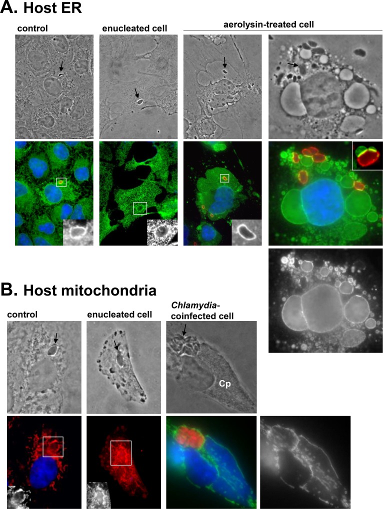
(A–B) Fluorescence microscopy of host ER with anti-calnexin antibody (green in [A]) and host mitochondria with mito-Tracker (red in B) or anti-Tom20 antibody (green in [B]). Nuclei are DAPI-stained. (A) Host ER association with the PV (identified by arrows on phase-contrast images or stained with anti-GRA7 antibody in red) in control mammalian cells, enucleated cells, or cells treated with aerolysin, which induces the vacuolization of host ER [41]. For this last condition, Vero cells were preincubated with 0.38-nM aerolysin for 2 h and infected for 10 min without toxin, then re-exposed to 0.38-nM aerolysin for 30 min. Despite extensive deformation of the host ER with the toxin, the parasite is able to attract this organelle, as exemplified by the intense zones of contact between the PV and host ER “bubbles,” clearly visible in the inset from another aerolysin-treated cell. (B) Host mitochondria association with the PV (identified by arrows on phase-contrast images) in control mammalian cells, enucleated cells, or cells coinfected with RFP-Toxoplasma (arrow) and Chlamydia psittaci (Cp) for 24 h. C. psittaci is notorious for attracting host mitochondria to its inclusion [42]. These bacteria multiply faster than Toxoplasma, occupying a large portion of the host cytoplasm. Despite these physical constraints, Toxoplasma manages to attract host mitochondria to the PV to a similar extent as Chlamydia. anti-Tom20, mitochondrial translocase of outer membrane; Cp, Chlamydia psittaci; GRA7, Toxoplasma parasitophorous vacuole membrane protein; ER, endoplasmic reticulum; PV, parasitophorous vacuole; RFP, red fluorescent protein.
References
-
- Sibley LD, Niesman IR, Parmley SF, Cesbron-Delauw MF. Regulated secretion of multi-lamellar vesicles leads to formation of a tubulo-vesicular network in host-cell vacuoles occupied by Toxoplasma gondii. J Cell Sci. 1995;108 (Pt 4): 1669–1677. - PubMed
-
- Magno RC, Lemgruber L, Vommaro RC, De Souza W, Attias M. Intravacuolar network may act as a mechanical support for Toxoplasma gondii inside the parasitophorous vacuole. Microsc Res Tech. 2005;67: 45–52. doi: 10.1002/jemt.20182 - DOI - PubMed
-
- De Souza W, Attias M. New views of the Toxoplasma gondii parasitophorous vacuole as revealed by Helium Ion Microscopy (HIM). J Struct Biol. 2015;191: 76–85. doi: 10.1016/j.jsb.2015.05.003 - DOI - PubMed
-
- Mercier C, Dubremetz J-F, Rauscher B, Lecordier L, Sibley LD, Cesbron-Delauw M-F. Biogenesis of nanotubular network in Toxoplasma parasitophorous vacuole induced by parasite proteins. Mol Biol Cell. 2002;13: 2397–2409. doi: 10.1091/mbc.E02-01-0021 - DOI - PMC - PubMed
-
- Caffaro CE, Boothroyd JC. Evidence for host cells as the major contributor of lipids in the intravacuolar network of Toxoplasma-infected cells. Eukaryotic Cell. 2011;10: 1095–1099. doi: 10.1128/EC.00002-11 - DOI - PMC - PubMed
Publication types
MeSH terms
Substances
Grants and funding
LinkOut - more resources
Full Text Sources
Other Literature Sources
Medical
