. 2018 May 4;46(8):4286-4300.
doi: 10.1093/nar/gky217.

A structurally conserved motif in γ-herpesvirus uracil-DNA glycosylases elicits duplex nucleotide-flipping

Affiliations

A structurally conserved motif in γ-herpesvirus uracil-DNA glycosylases elicits duplex nucleotide-flipping

Christopher Earl et al. Nucleic Acids Res. .

Abstract

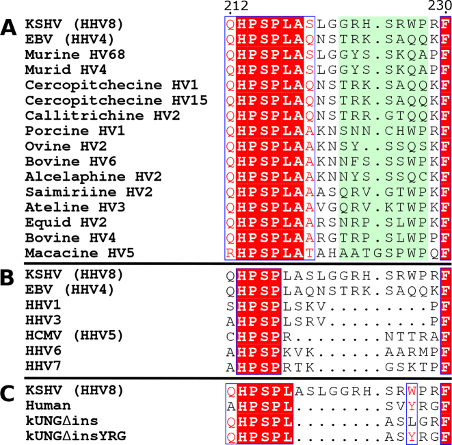
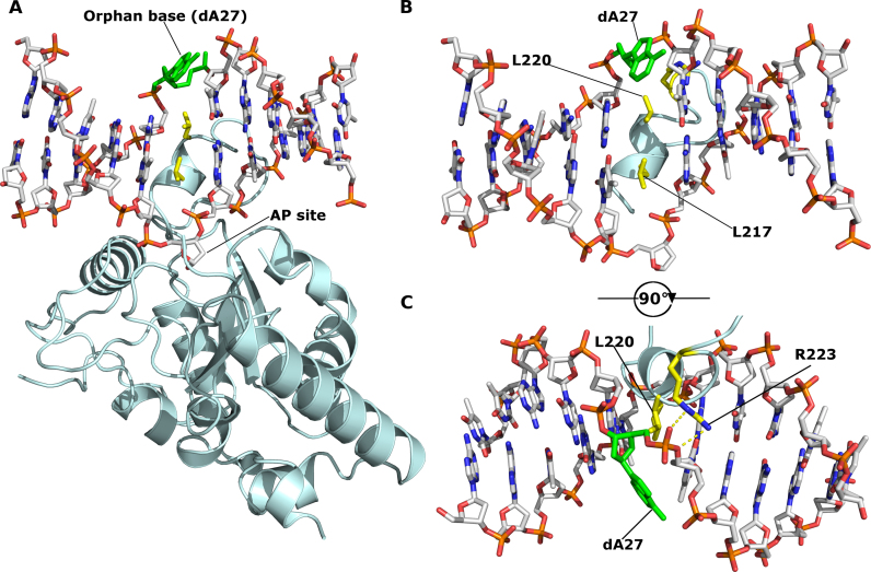
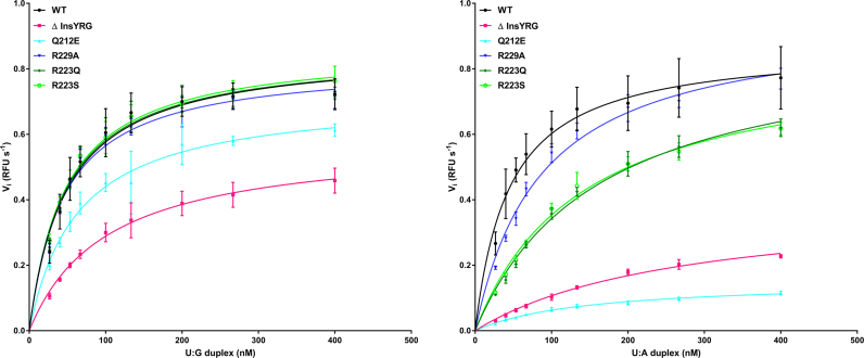
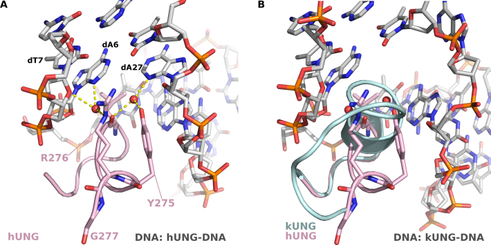
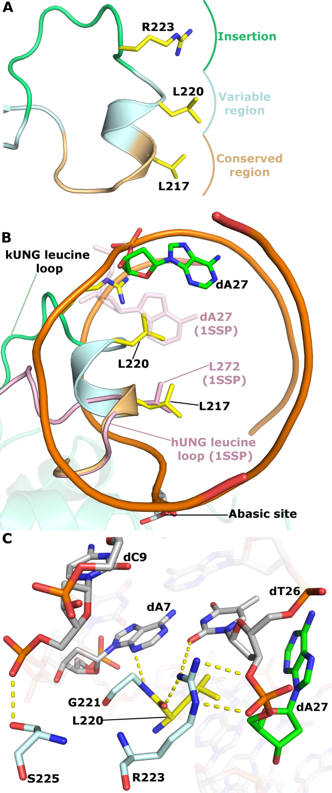
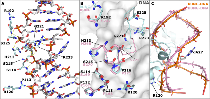
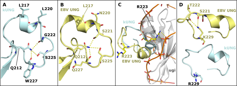
Efficient γ-herpesvirus lytic phase replication requires a virally encoded UNG-type uracil-DNA glycosylase as a structural element of the viral replisome. Uniquely, γ-herpesvirus UNGs carry a seven or eight residue insertion of variable sequence in the otherwise highly conserved minor-groove DNA binding loop. In Epstein-Barr Virus [HHV-4] UNG, this motif forms a disc-shaped loop structure of unclear significance. To ascertain the biological role of the loop insertion, we determined the crystal structure of Kaposi's sarcoma-associated herpesvirus [HHV-8] UNG (kUNG) in its product complex with a uracil-containing dsDNA, as well as two structures of kUNG in its apo state. We find the disc-like conformation is conserved, but only when the kUNG DNA-binding cleft is occupied. Surprisingly, kUNG uses this structure to flip the orphaned partner base of the substrate deoxyuridine out of the DNA duplex while retaining canonical UNG deoxyuridine-flipping and catalysis. The orphan base is stably posed in the DNA major groove which, due to DNA backbone manipulation by kUNG, is more open than in other UNG-dsDNA structures. Mutagenesis suggests a model in which the kUNG loop is pinned outside the DNA-binding cleft until DNA docking promotes rigid structuring of the loop and duplex nucleotide flipping, a novel observation for UNGs.


Figures

Figure 1.
Figure 1.
Sequence alignments of UNG leucine loops, adapted from ESPript web server outputs (19). Within each alignment, strictly conserved residues are written in white and highlighted in red. Residues with >70% conservation are surrounded by a blue box and written in red. (A) Leucine loop sequences of γ-herpesviruses with leucine loop extensions highlighted in pale green. (B) Leucine loop sequences of all known human herpesviruses. (C) Leucine loop sequences of kUNG, human UNG and kUNG mutants used in this study.
Figure 2.
Figure 2.
kUNG (pale cyan cartoon) in complex with DNA (sticks with grey carbons). Labelled residues, discussed below, are displayed as yellow sticks. The orphan base, dA27, is shown as green sticks. The AP site formed after uracil cleavage is labelled, no density was present for the cleaved uracil. (A) The overall architecture of the kUNG–dsDNA complex. (B and C) View of DNA and the kUNG leucine loop, only DNA and leucine loop residues are shown. Hydrogen bonds/electrostatic interactions are shown as yellow dashes. The leucine ‘loop’ includes a helical region that invades the minor groove presenting two leucine residues: L217, the canonical ‘doorstop’ residue which displaces the substrate uracil, and L220 which displaces the opposing ‘orphan’ base, dA27. The adenine base of dA27 is flipped out of the DNA duplex into the exterior environment, this is accompanied by the support of a strained backbone conformation by interactions with R223 of kUNG.
Figure 3.
Figure 3.
Relative enzymatic characteristics of wild-type kUNG, and mutants prepared for this study; 6.65 nM kUNG variant per assay: wild-type (black line, solid circle points), ΔinsYRG (pink line, solid square points), Q212E (cyan line, triangle points), R223Q (dark green line, diamond points), R223S (bright green line, hollow circle points), R229A (dark blue line, inverted triangle points). The substrate concentration is along the x-axis, and RFU s−1 is along the y-axis. (A) Substrate duplex DNA contains a U:G mismatch. (B) Substrate duplex DNA contains a U:A pair. A minimum of four measurements per duplex DNA series were performed; see text for full details.
Figure 4.
Figure 4.
(A) Interactions between the hUNG ‘minor groove reading head’ and the DNA minor groove (PDB code: 1SSP). The hUNG leucine loop is shown as a pink cartoon with the Y275, R276 and G277 shown as sticks. DNA is shown in sticks with grey carbons, water molecules are shown as red spheres, hydrogen-bonds are shown as yellow dashes. R276 forms water-mediated hydrogen bonds with the adenine base of dA6 and the deoxyribose moiety of dT7, the nucleotide immediately 3′ of the uracil. Y275 forms a water-mediated hydrogen bond with the adenine base of dA27, the ‘orphan’ base. These interactions serve to widen the minor groove allowing the leucine loop to enter and perform nucleotide-flipping. (B) View [as in panel A] of the kUNG–dsDNA structure showing the kUNG leucine loop in pale cyan cartoon. The hUNG leucine loop is included [as in panel A]. The architecture of the kUNG leucine loop in this region is entirely different to that of hUNG with the kUNG protein backbone being inserted into the DNA duplex.
Figure 5.
Figure 5.
kUNG leucine loop structure. (A) Schematic of the conserved, variable and extension regions of the kUNG leucine loop. The strictly conserved ‘doorstop’ leucine L217, orphan nucleotide-flipping leucine L220 and key DNA-binding residue R223 sidechains are shown as sticks with yellow carbons. (B) View along the axis of the DNA double helix comparing kUNG and hUNG leucine loop positions, the DNA backbone is shown as an orange ribbon, most nucleotides are omitted for clarity. The kUNG leucine loop is coloured [as in panel A], the hUNG leucine loop, from PDB code: 1SSP (31), is shown in pink cartoon representation with the ‘doorstop’ leucine shown as pink sticks. The orphan dA27 nucleotide in the kUNG–dsDNA structure is shown in sticks with green carbons. The orphan base in 1SSP (faded pink sticks) takes its usual position in the base stack; this position would be precluded by steric hindrance from L220 of kUNG. (C) Stick representation of key protein–DNA interactions between the variable/extension regions of the leucine loop and the DNA minor groove. Protein residues are shown as sticks with pale cyan carbons, L220 is shown with yellow carbons. DNA is coloured [as in panel B]. Hydrogen bonds/electrostatic interactions are shown as yellow dashes. The guanidinium head group of R223 interacts with the phosphate group of dA27 and O2 of the dT26 base, as well as the carbonyl oxygen of L220. Further interactions from the kUNG leucine loop in the minor groove come from S225 and G221.
Figure 6.
Figure 6.
Protein–DNA contacts in the kUNG–dsDNA complex, see Supplementary Table S1 for details. (A) Stick representation showing DNA-contacting kUNG residues (pale cyan carbons), all other protein residues are omitted for clarity. Hydrogen bonds/electrostatic interactions are shown as yellow dashes. (B) Alignment of kUNG–dsDNA and hUNG–dsDNA (PDB code: 1SSP) complexes displaying structural conservation of the ‘Ser-Pro pinch' residues. kUNG residues are shown as sticks with pale cyan carbons, hUNG residues are shown as sticks with pink carbons. kUNG and hUNG leucine loop backbones are shown as ribbons in pale cyan and pink respectively. DNA from the kUNG–dsDNA complex is shown as a grey surface. Despite excellent overall conservation, the action of S273 in hUNG as a hydrogen bond donor is not mimicked by kUNG. Instead, contributions from G221, R223 and S225 provide ‘pinching’ interactions to compress the DNA backbone. R223 of kUNG provides an additional contact with the DNA backbone not seen in hUNG. (C) Comparison of the global DNA backbone conformation in enzyme–product complexes of kUNG (orange DNA) and hUNG (pink DNA). DNA backbones are shown as ribbon traces between phosphates. 3′ of the uracil (at the top of the image), the DNA backbone position is largely similar. 5′ of the uracil however, the DNA takes up a position closer to kUNG than in hUNG, a position supported by contact between R223 and the phosphate of dA29. There is a less pronounced kink in the DNA by kUNG than is seen in hUNG–dsDNA structures. Consequently, the flipped out orphan base dA27 is presented to the solvent in a more accessible major groove.
Figure 7.
Figure 7.
Comparison of leucine loop structures in the kUNG–dsDNA and EBV UNG–Ugi complexes (PDB code: 2J8X). (A and B) The leucine loops of kUNG (A, pale cyan) and EBV UNG (B, yellow) have similar structures including a rigid conformation of the extension region being centred on hydrogen-bonding with Q212. Residues involved in this hydrogen-bonding network and nucleotide-flipping residues are shown as sticks. Hydrogen bonds/electrostatic interactions are shown as yellow dashes. (C) Relative positions of the kUNG and EBV UNG leucine loops with the two UNGs aligned on conserved catalytic core residues. The kUNG leucine loop (pale cyan) invades the minor groove of the DNA (shown as an orange trace between phosphates). The EBV UNG leucine loop (yellow) is precluded from the same position as the kUNG loop by the bulky nature of Ugi (grey cartoon). R223 forms protein–DNA contacts in kUNG but protrudes into the solvent in the Ugi-bound EBV UNG structure. (D) Relative positions of R/K229. In the EBV UNG–Ugi structure (yellow), the side-chain of K229 is oriented towards the centre of the loop and interacts with T222 and S221. The side-chain of R229 of kUNG (pale cyan) forms no contacts with other atoms. R/K229 may act as a pre-catalytic ‘pinning’ residue to hold the loop away from the DNA binding cleft to allow DNA binding.

Similar articles

Cited by

References

    1. Cesarman E., Chang Y., Moore P.S., Said J.W., Knowles D.M.. Kaposi's sarcoma–associated herpesvirus-like dna sequences in aids-related body-cavity–based lymphomas e. N. Engl. J. Med. 1995; 332:1186–1191. - PubMed
    1. Soulier J., Grollet L., Oksenhendler E., Cacoub P., Cazals-Hatem D., Babinet P., d’Agay M.F., Clauvel J.P., Raphael M., Degos L.. Kaposi's sarcoma-associated herpesvirus-like DNA sequences in multicentric Castleman's disease. Blood. 1995; 86:1276–1280. - PubMed
    1. Arvanitakis L., Mesri E.A., Nador R.G., Said J.W., Asch A.S., Knowles D.M., Cesarman E.. Establishment and characterization of a primary effusion (body cavity-based) lymphoma cell line (BC-3) harboring kaposi's sarcoma-associated herpesvirus (KSHV/HHV-8) in the absence of Epstein-Barr virus. Blood. 1996; 88:2648–2654. - PubMed
    1. Parravicini C., Chandran B., Corbellino M., Berti E., Paulli M., Moore P.S., Chang Y.. Differential Viral Protein Expression in Kaposi's Sarcoma-Associated Herpesvirus-Infected Diseases. Am. J. Pathol. 2000; 156:743–749. - PMC - PubMed
    1. Paulose-Murphy M., Ha N., Xiang C., Chen Y., Gillim L., Yarchoan R., Meltzer P., Bittner M., Trent J., Zeichner S.. Transcription program of human herpesvirus 8 (Kaposi's sarcoma-associated herpesvirus). J. Virol. 2001; 75:4843–4853. - PMC - PubMed

Publication types

LinkOut - more resources