Review
. 2018 Mar 28;7(4):26.
doi: 10.3390/cells7040026.

Microglia after Seizures and in Epilepsy

Affiliations
Review

Microglia after Seizures and in Epilepsy

Toshimitsu Hiragi et al. Cells. .

Abstract

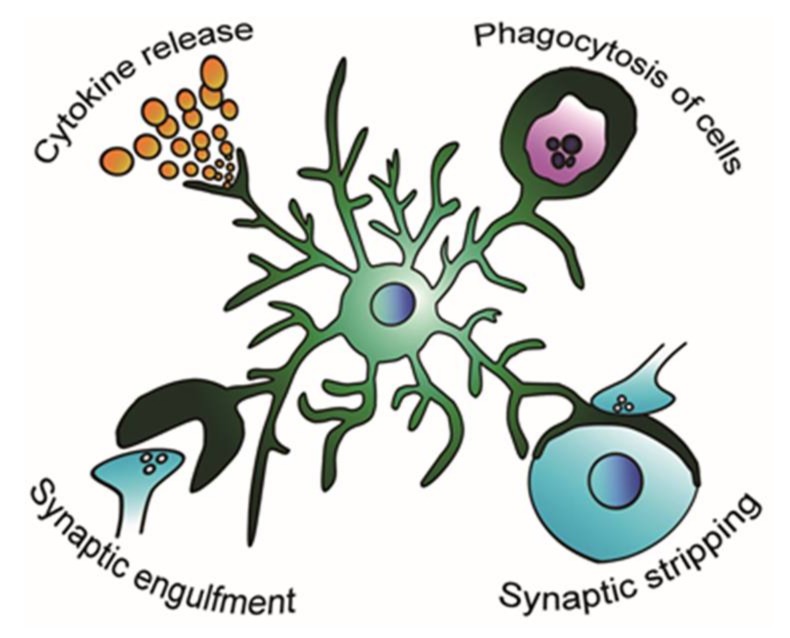
Microglia are the resident immune cells in the brain that constitute the brain's innate immune system. Recent studies have revealed various functions of microglia in the development and maintenance of the central nervous system (CNS) in both health and disease. However, the role of microglia in epilepsy remains largely undiscovered, partly because of the complex phenotypes of activated microglia. Activated microglia likely exert different effects on brain function depending on the phase of epileptogenesis. In this review, we mainly focus on the animal models of temporal lobe epilepsy (TLE) and discuss the proepileptic and antiepileptic roles of activated microglia in the epileptic brain. Specifically, we focus on the roles of microglia in the production of inflammatory cytokines, regulation of neurogenesis, and surveillance of the surrounding environment in epilepsy.

Keywords: epilepsy; epileptogenesis; glia; microglia; seizure; synapse.


Conflict of interest statement

The authors declare no conflicts of interest.

Figures

Figure 1
Figure 1
Possible modification of the microglial role in the epileptic brain. Microglia respond to the epileptic environment and change their activity of releasing proinflammatory and anti-inflammatory cytokines [16], phagocytosing apoptotic and living cells [18,19], engulfing synapses [20], and stripping synapses [21].

References

    1. Cunningham C.L., Martinez-Cerdeno V., Noctor S.C. Microglia Regulate the Number of Neural Precursor Cells in the Developing Cerebral Cortex. J. Neurosci. 2013;33:4216–4233. doi: 10.1523/JNEUROSCI.3441-12.2013. - DOI - PMC - PubMed
    1. Sierra A., Encinas J.M., Deudero J.J.P., Chancey J.H., Enikolopov G., Overstreet-Wadiche L.S., Tsirka S.E., Maletic-Savatic M. Microglia shape adult hippocampal neurogenesis through apoptosis-coupled phagocytosis. Cell Stem Cell. 2010;7:483–495. doi: 10.1016/j.stem.2010.08.014. - DOI - PMC - PubMed
    1. Ueno M., Fujita Y., Tanaka T., Nakamura Y., Kikuta J., Ishii M., Yamashita T. Layer V cortical neurons require microglial support for survival during postnatal development. Nat. Neurosci. 2013;16:543–551. doi: 10.1038/nn.3358. - DOI - PubMed
    1. Peri F., Nüsslein-Volhard C. Live Imaging of Neuronal Degradation by Microglia Reveals a Role for v0-ATPase a1 in Phagosomal Fusion in Vivo. Cell. 2008;133:916–927. doi: 10.1016/j.cell.2008.04.037. - DOI - PubMed
    1. Svahn A.J., Graeber M.B., Ellett F., Lieschke G.J., Rinkwitz S., Bennett M.R., Becker T.S. Development of ramified microglia from early macrophages in the zebrafish optic tectum. Dev. Neurobiol. 2013;73:60–71. doi: 10.1002/dneu.22039. - DOI - PubMed

LinkOut - more resources