

Exosomes from Endothelial Progenitor Cells Improve the Outcome of a Murine Model of Sepsis
- PMID: 29599080
- PMCID: PMC5993985
- DOI: 10.1016/j.ymthe.2018.02.020
Exosomes from Endothelial Progenitor Cells Improve the Outcome of a Murine Model of Sepsis
Abstract
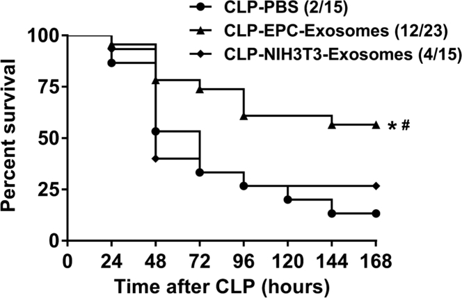
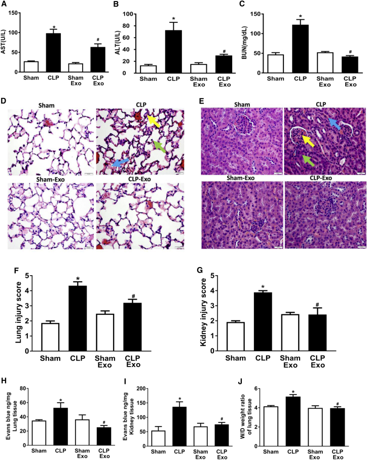
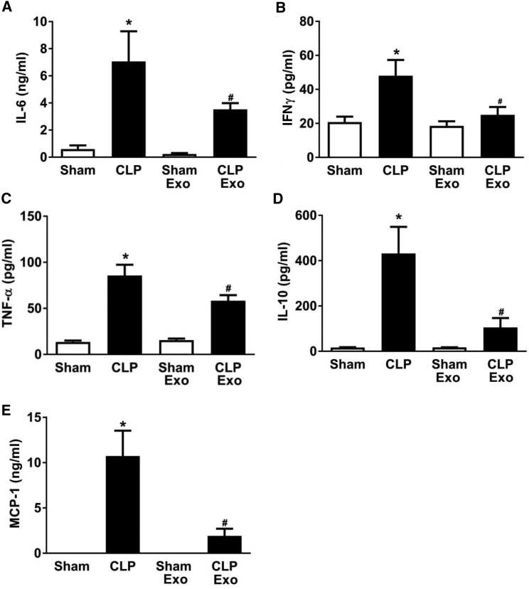
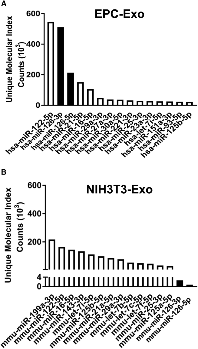
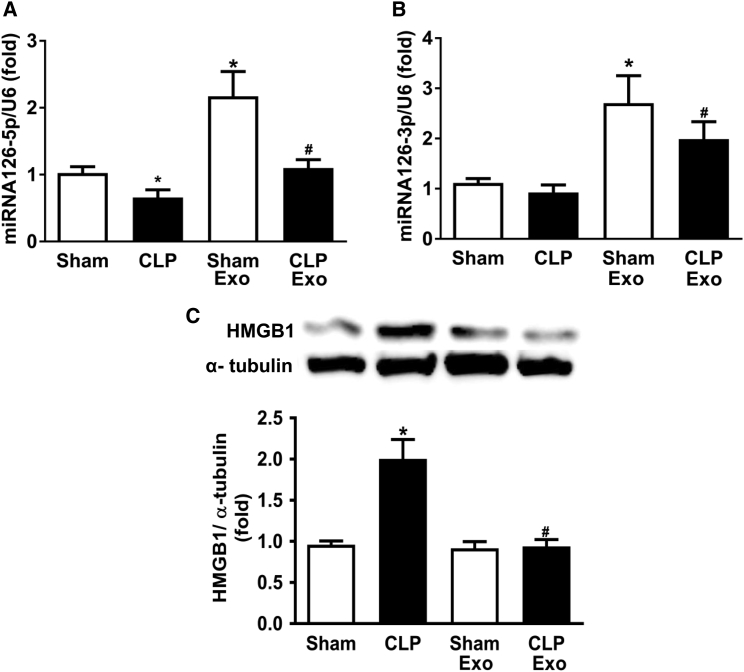
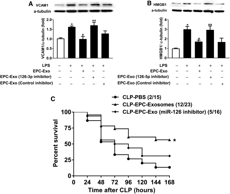
Microvascular dysfunction leads to multi-organ failure and mortality in sepsis. Our previous studies demonstrated that administration of exogenous endothelial progenitor cells (EPCs) confers protection in sepsis as evidenced by reduced vascular leakage, improved organ function, and increased survival. We hypothesize that EPCs protect the microvasculature through the exosomes-mediated transfer of microRNAs (miRNAs). Mice were rendered septic by cecal ligation and puncture (CLP), and EPC exosomes were administered intravenously at 4 hr after CLP. EPC exosomes treatment improved survival, suppressing lung and renal vascular leakage, and reducing liver and kidney dysfunction in septic mice. EPC exosomes attenuated sepsis-induced increases in plasma levels of cytokines and chemokine. Moreover, we determined miRNA contents of EPC exosomes with next-generation sequencing and found abundant miR-126-3p and 5p. We demonstrated that exosomal miR-126-5p and 3p suppressed LPS-induced high mobility group box 1 (HMGB1) and vascular cell adhesion molecule 1 (VCAM1) levels, respectively, in human microvascular endothelial cells (HMVECs). Inhibition of microRNA-126-5p and 3p through transfection with microRNA-126-5p and 3p inhibitors abrogated the beneficial effect of EPC exosomes. The inhibition of exosomal microRNA-126 failed to block LPS-induced increase in HMGB1 and VCAM1 protein levels in HMVECs and negated the protective effect of exosomes on sepsis survival. Thus, EPC exosomes prevent microvascular dysfunction and improve sepsis outcomes potentially through the delivery of miR-126.
Keywords: barrier function; endothelial progenitor cells; exosomes; microRNA; sepsis.
Copyright © 2018 The American Society of Gene and Cell Therapy. Published by Elsevier Inc. All rights reserved.
Figures
Similar articles
-
Endothelial progenitor cells-secreted extracellular vesicles containing microRNA-93-5p confer protection against sepsis-induced acute kidney injury via the KDM6B/H3K27me3/TNF-α axis.Exp Cell Res. 2020 Oct 15;395(2):112173. doi: 10.1016/j.yexcr.2020.112173. Epub 2020 Jul 15. Exp Cell Res. 2020. PMID: 32679234
-
Exosomes from endothelial progenitor cells improve outcomes of the lipopolysaccharide-induced acute lung injury.Crit Care. 2019 Feb 13;23(1):44. doi: 10.1186/s13054-019-2339-3. Crit Care. 2019. PMID: 30760290 Free PMC article.
-
Endothelial progenitor cells and a stromal cell-derived factor-1α analogue synergistically improve survival in sepsis.Am J Respir Crit Care Med. 2014 Jun 15;189(12):1509-19. doi: 10.1164/rccm.201312-2163OC. Am J Respir Crit Care Med. 2014. PMID: 24707934 Free PMC article.
-
MicroRNA-126 Regulates Thrombosis Through Endothelial Progenitor Cells.DNA Cell Biol. 2023 Jun;42(6):315-321. doi: 10.1089/dna.2022.0643. Epub 2023 Apr 10. DNA Cell Biol. 2023. PMID: 37036794 Review.
-
Mobilization of endothelial progenitor cells in sepsis.Inflamm Res. 2020 Jan;69(1):1-9. doi: 10.1007/s00011-019-01299-9. Epub 2019 Nov 22. Inflamm Res. 2020. PMID: 31758219 Review.
Cited by
-
Pharmacologic therapies of ARDS: From natural herb to nanomedicine.Front Pharmacol. 2022 Oct 28;13:930593. doi: 10.3389/fphar.2022.930593. eCollection 2022. Front Pharmacol. 2022. PMID: 36386221 Free PMC article. Review.
-
The Role of MicroRNAs in Acute Respiratory Distress Syndrome and Sepsis, From Targets to Therapies: A Narrative Review.Anesth Analg. 2020 Nov;131(5):1471-1484. doi: 10.1213/ANE.0000000000005146. Anesth Analg. 2020. PMID: 33079870 Free PMC article. Review.
-
Exosomes derived from endothelial progenitor cells ameliorate glyoxylate deprivation (OGD)-induced neuronal apoptosis by delivering miR-221-3p.Histol Histopathol. 2023 Apr;38(4):423-430. doi: 10.14670/HH-18-528. Epub 2022 Oct 3. Histol Histopathol. 2023. PMID: 36190183
-
Endothelial colony-forming cell-derived exosomal miR-21-5p regulates autophagic flux to promote vascular endothelial repair by inhibiting SIPL1A2 in atherosclerosis.Cell Commun Signal. 2022 Mar 12;20(1):30. doi: 10.1186/s12964-022-00828-0. Cell Commun Signal. 2022. PMID: 35279183 Free PMC article.
-
Human Amnion Epithelial Cells and Their Derived Exosomes Alleviate Sepsis-Associated Acute Kidney Injury via Mitigating Endothelial Dysfunction.Front Med (Lausanne). 2022 Mar 24;9:829606. doi: 10.3389/fmed.2022.829606. eCollection 2022. Front Med (Lausanne). 2022. PMID: 35402422 Free PMC article.
References
-
- Freund Y., Lemachatti N., Krastinova E., Van Laer M., Claessens Y.E., Avondo A., Occelli C., Feral-Pierssens A.L., Truchot J., Ortega M., French Society of Emergency Medicine Collaborators Group Prognostic accuracy of sepsis-3 criteria for in-hospital mortality among patients with suspected infection presenting to the emergency department. JAMA. 2017;317:301–308. - PubMed
-
- Shankar-Hari M., Phillips G.S., Levy M.L., Seymour C.W., Liu V.X., Deutschman C.S., Angus D.C., Rubenfeld G.D., Singer M., Sepsis Definitions Task Force Developing a new definition and assessing new clinical criteria for septic shock: for the Third International Consensus Definitions for Sepsis and Septic Shock (Sepsis-3) JAMA. 2016;315:775–787. - PMC - PubMed
-
- Rudd K.E., Delaney A., Finfer S. Counting sepsis, an imprecise but improving science. JAMA. 2017;318:1228–1229. - PubMed
-
- Doerschug K.C., Delsing A.S., Schmidt G.A., Haynes W.G. Impairments in microvascular reactivity are related to organ failure in human sepsis. Am. J. Physiol. Heart Circ. Physiol. 2007;293:H1065–H1071. - PubMed
Publication types
MeSH terms
Substances
Grants and funding
LinkOut - more resources
Full Text Sources
Other Literature Sources
Medical
Miscellaneous
