. 2018 Jun;38(6):1333-1345.
doi: 10.1161/ATVBAHA.118.310951. Epub 2018 Mar 29.

CaMKII (Ca2+/Calmodulin-Dependent Kinase II) in Mitochondria of Smooth Muscle Cells Controls Mitochondrial Mobility, Migration, and Neointima Formation

Affiliations

CaMKII (Ca2+/Calmodulin-Dependent Kinase II) in Mitochondria of Smooth Muscle Cells Controls Mitochondrial Mobility, Migration, and Neointima Formation

Emily K Nguyen et al. Arterioscler Thromb Vasc Biol. 2018 Jun.

Abstract

Objective: The main objective of this study is to define the mechanisms by which mitochondria control vascular smooth muscle cell (VSMC) migration and impact neointimal hyperplasia.

Approach and results: The multifunctional CaMKII (Ca2+/calmodulin-dependent kinase II) in the mitochondrial matrix of VSMC drove a feed-forward circuit with the mitochondrial Ca2+ uniporter (MCU) to promote matrix Ca2+ influx. MCU was necessary for the activation of mitochondrial CaMKII (mtCaMKII), whereas mtCaMKII phosphorylated MCU at the regulatory site S92 that promotes Ca2+ entry. mtCaMKII was necessary and sufficient for platelet-derived growth factor-induced mitochondrial Ca2+ uptake. This effect was dependent on MCU. mtCaMKII and MCU inhibition abrogated VSMC migration and mitochondrial translocation to the leading edge. Overexpression of wild-type MCU, but not MCU S92A, mutant in MCU-/- VSMC rescued migration and mitochondrial mobility. Inhibition of microtubule, but not of actin assembly, blocked mitochondrial mobility. The outer mitochondrial membrane GTPase Miro-1 promotes mitochondrial mobility via microtubule transport but arrests it in subcellular domains of high Ca2+ concentrations. In Miro-1-/- VSMC, mitochondrial mobility and VSMC migration were abolished, and overexpression of mtCaMKII or a CaMKII inhibitory peptide in mitochondria (mtCaMKIIN) had no effect. Consistently, inhibition of mtCaMKII increased and prolonged cytosolic Ca2+ transients. mtCaMKII inhibition diminished phosphorylation of focal adhesion kinase and myosin light chain, leading to reduced focal adhesion turnover and cytoskeletal remodeling. In a transgenic model of selective mitochondrial CaMKII inhibition in VSMC, neointimal hyperplasia was significantly reduced after vascular injury.

Conclusions: These findings identify mitochondrial CaMKII as a key regulator of mitochondrial Ca2+ uptake via MCU, thereby controlling mitochondrial translocation and VSMC migration after vascular injury.

Keywords: calcium; calcium-calmodulin-dependent protein kinase type 2; hyperplasia; microtubules; mitochondria; neointima.


Conflict of interest statement

Disclosures: One author is a named inventor on awarded patents related to targeting CaMKII inhibitors to mitochondria (OMK). All other authors declare no conflict of interest exists

Figures

Figure 1
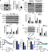
Figure 1. Mitochondrial CaMKII regulates a feed forward circuit promoting mitochondrial Ca2+ uptake
A) Representative immunoblot and summary data for CaMKII in mitochondrial (mito) and cytosolic (cyto) fractions and mitoplasts from VSMC. Mitoplasts (inner mitochondrial membrane and matrix) were generated by digestion of mitochondrial fractions with Proteinase K. GAPDH: cytosolic marker; Cox IV: marker of inner mitochondrial membrane; Drp1: marker of outer mitochondrial membrane. Data quantified as intensity of CaMKII to total amount of protein by BCA protein assay (n=4 independent experiments), *p<0.05 versus cytosolic fraction by Kruskall-Wallis. B) Immunoblot for active (phosphorylated, pCaMKII) and total CaMKII in whole cell lysate and mitoplast fractions of VSMC from MCU−/− or WT mice before or after treatment with PDGF (20 ng/mL) for 15 min after serum starvation (SF). (n=5 independent experiments). C) Immunoblot for pCaMKII in mitoplast fractions of MCU−/− VSMC after transfection of WT MCUor MCU mutant Ser92 to Ala (S92A) or control and in mitoplasts of WT VSMC; treated as in (B).(n=5 independent experiments). D) Active (phosphorylated, pMCU, at S92) and total MCU inmitochondrial fractions from VSMC expressing mtCaMKIIN or control. COX IV: loading control. (n=5 independent experiments). For B-D, *p<0.05 versus control SF, #p<0.05 versus control PDGF by 2-way ANOVA. E) Representative mtPericam tracing in VSMC expressing mtCaMKIIN or control for 48 hr; arrow indicates addition of PDGF (20 ng/mL). Peak amplitude of mtPericam response normalized to peak amplitude in control cells and area under the curve (AUC) of mtPericam signal in 15 cells per condition (n=3 independent experiments). F) Ca2+ concentration by o-cresolphthalein assay in mitochondria isolated from VSMC expressing mtCaMKIIN or control for 48 hr; data were normalized to total mitochondrial protein content (n=5). G) Representative mtPericam tracing in WT or MCU −/− VSMC expressing mtCaMKII or control for 48 hr with addition of PDGF, *p<0.05 versus control by Student’s t-test.
Figure 2
Figure 2. Mitochondrial CaMKII inhibition blocks VSMC migration
A) Boyden chamber migration assay in VSMC expressing mtCaMKIIN (MOI 50) or control in SF media or after treatment with PDGF (20 ng/mL) for 6 hr (n=5 independent experiments). B) Scratch wound assay in VSMC expressing mtCaMKIIN or control in SF media or after treatment with PDGF (20 ng/mL); data quantified as percent wound closure at 24 hr compared to 0 hr (n=5 independent experiments). C) Representative DPC images from high throughput scratch wound assay. WT VSMC were exposed to increasing MOIs of Ad-mtCaMKIIN; D) Migration in C quantified as percent wound closure at 24 hr compared to 0 hr; *p<0.05 versus MOI of 0 by 1-way ANOVA. E) Scratch wound assay in VSMC expressing mtCaMKII or control after treatment with PDGF; data quantified as percent wound closure at 24 hr as compared to 0 hr. *p<0.05 by Student’s T-test. F) High throughput scratch wound assay in MCU−/− or WT VSMC with expression of mtCaMKIIN or mtCaMKII after treatment with PDGF. High throughput experiments were conducted in quadruplicate, with biological replicates of three independent experiments. For A, B, and F, *p<0.05 versus control in SF, #p<0.05 versus PDGF-treated control by 2-way ANOVA.
Figure 3
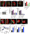
Figure 3. Mitochondrial CaMKII inhibition blocks translocation of mitochondria to the leading edge of migrating VSMC
A) Representative images of mitochondrial localization after scratch wound and PDGF treatment (20 ng/mL) at indicated time points; green: Ad-mtGFP; red: phalloidin-568. B) Quantification of mitochondrial distribution by line scan analysis in the leading to anterior cell areas and quantification of mitochondrial distribution expressed as percent mtGFP signal in the leading cell area (region of interest/ROI) relative to total mtGFP signal per cell (see details in methods and Supplemental Figure 5D); *p<0.05 versus control at 0 hr by 1-way ANOVA. C) Representative images of mitochondrial localization in migrating VSMC before and after PDGF treatment. Studies were performed in WT VSMC expressing mtCaMKIIN or control, MCU−/− VSMC, or WT VSMC treated with Ru360 (1 µM) or mitoTEMPO (10 µM). mitochondrial distribution was quantified by line scan analysis. D) Representative images of mitochondrial localization in migrating MCU−/− VSMC transfected with control plasmid, WT MCU, or S92A MCU, before and after PDGF. Mitochondrial distribution was quantified by line scan analysis in the leading to anterior cell areas and expressed as percent mtGFP signal in the leading cell. Analyses were performed on 50 cells per condition (n=3 independent experiments). For C and D, *p<0.05 versus control at 0 hr, #p<0.05 versus control at the same time point by 2-way ANOVA. Scale bar= 10 µm. E) Scratch wound assay in cells from D; data quantified as percent wound closure at 6 hr after scratch as compared to 0 hr (n=3 independent experiments). *p<0.05 versus control by 1-way ANOVA.
Figure 4
Figure 4. mtCaMKII regulation of VSMC migration and mitochondrial mobility is dependent on the Ca2+-sensitive Miro-1
A) Representative images of mitochondrial localization in migrating VSMC treated for 6 hr with PDGF and either actin inhibitor Cytochalasin D (0.1nM) or microtubule inhibitor Nocodazole (1 µM) or Nocodazole washed out 1 hr before fixation. Quantification of mitochondrial distribution expressed as percent mtGFP signal in the leading cell area by line scan. * p<0.05 vs vehicle, # p<0.05 vs Nocodazole by 1-way ANOVA. (20 cells over 3 independent experiments) B) Representative immunoblot of Miro-1 expression in Miro fl/fl VSMC with and without Ad5-Cre infection (MOI 10). C) VSMC migration by traditional scratch wound assay in Mirofl/fl or Cre-infected Mirofl/fl (Miro−/−) VSMC co-expressing mtCaMKII or mtCaMKIIN. Data quantified as percent wound closure at 24 hr as compared to 0 hr, (n = 3 independent experiments). D) Quantification of mitochondrial distribution by line scan analysis in the leading to anterior cell areas expressed as percent mtGFP signal in the leading cell (30 cells over 3 independent experiments). E) Representative images of mitochondrial localization after scratch wound and PDGF treatment (20 ng/mL) at 24 hr after scratch. Green: mitochondria, AdmtGFP; red: F-actin, phalloidin-568. For A-D, *p<0.05 versus control Mirofl/fl by 1-way ANOVA. Scale bar = 10µm. F) Representative cytosolic Ca2+ tracing by Fura-2AM after 20 ng/ml PDGF (arrow) in VSMC expressing control or mtCaMKIIN. Quantification of Fura-2AM signal as time constant of decay (τ, left), residual signal at 300 s after PDGF relative to peak response (middle) and AUC of Fura-2AM signal (right) in 25 cells per condition in 5 independent experiments. *p<0.05 versus control by Student’s T-test.
Figure 5
Figure 5. Mitochondrial CaMKII inhibition alters focal adhesions dynamics
A) Immunofluorescent overlaid time-lapse images of FA (GFP-Vinculin) at 0 (green) and 90 (red) min in VSMC expressing mtCaMKIIN or control, and in the presence of 20 ng/mL PDGF. FA colocalization at 0 and 90 min was quantified in 15 cells using CellProfiler software, *p<0.05 vs SF, #p<0.05 vs control PDGF by 1-way ANOVA. B) Representative images of focal adhesion (FA) at wound edge in VSMC expressing mtCaMKIIN or control at 0 or 6 hr after PDGF treatment in SF media with quantification of average focal adhesion size (red: phalloidin, actin;green: vinculin, FA marker). C) Immunoblots for phosphorylated (pFAK Y577) and total FAK in whole cell lysates from VSMC expressing mtCaMKIIN and treated with PDGF (20 ng/ml) for 0–60 min. Data were quantified as phosphorylated FAK (pFAK Y577) relative to total FAK, normalized to respective baseline at 0 min. GAPDH: loading control (n=4 independent experiments). For B and C, *p<0.05 versus control SF, #p<0.05 versus control PDGF by 2-way ANOVA. Scale bar= 10 µm.
Figure 6
Figure 6. Mitochondrial CaMKII inhibition in vivo blocks neointimal hyperplasia following endothelial injury
A) Representative images of carotid arteries from C57/Bl6 (WT) and SMmtCaMKIIN mice with and without tamoxifen (Tamox) treatment, which induces cre recombination. Green: ubiquitously expressed GFP; red: mtCaMKIIN; blue: Nuclei. “L” indicates arterial lumen; scale bar= 20 µm. B) Immunoblots for hemagglutinin (HA)-tagged CaMKIIN in mitochondrial or cytosolic fractions isolated from aortas of SM-mtCaMKIIN mice with and without tamoxifen treatment. VSMC infected with Ad-mtCaMKIIN aspositive control; Cox IV: mitochondrial marker. C) Ca2+ concentration by o-cresolphthalein assay in mitochondria isolated from aortas of SM-mtCaMKIIN or WT littermate controls (SM-Cre mice). Data were normalized to total mitochondrial protein measured by BCA assay (n=5 mice per genotype). D) Representative images of Verhoeff’s Van Gieson staining of left common carotid arteries at 28 days after endothelial injury in WT or SM-mtCaMKIIN mice. Contralateral (right) common carotid artery serves as uninjured control with quantification of neointimal area at 200 µm from the carotid bifurcation and neointimal volume calculated from all neointimal areas within 500 µm from carotid bifurcation; scale bar= 50 µm, n=12 mice per genotype. *p<0.05 versus WT by Student’s T-test. E) Graphic representation of the proposed pathway by which mitochondrial CaMKII regulation of mitochondrial Ca2+, mobility, and VSMC migration. mtCaMKII regulates a feed-forward circuit that promotes mitochondrial Ca2+ uptake. Inhibition of mtCaMKII leads to decreased mitochondrial Ca2+ uptake, leading to decreased Ca2+ clearance in the cytosol and mitochondrial arrest. This prevents mitochondria from translocating to focal adhesions, thus inhibiting VSMC migration and neointima formation.

Comment in

  • Smooth Muscle Cells Move With Mitochondria.
    Shi N, Chen SY. Shi N, et al. Arterioscler Thromb Vasc Biol. 2018 Jun;38(6):1255-1257. doi: 10.1161/ATVBAHA.118.311085. Arterioscler Thromb Vasc Biol. 2018. PMID: 29793991 Free PMC article. No abstract available.

Similar articles

Cited by

References

    1. Moses JW, Leon MB, Popma JJ, Fitzgerald PJ, Holmes DR, O'Shaughnessy C, Caputo RP, Kereiakes DJ, Williams DO, Teirstein PS, Jaeger JL, Kuntz RE Investigators S. Sirolimus-eluting stents versus standard stents in patients with stenosis in a native coronary artery. The New England journal of medicine. 2003;349:1315–1323. - PubMed
    1. Schwartz SM. Perspectives series: cell adhesion in vascular biology. Smooth muscle migration in atherosclerosis and restenosis. The Journal of Clinical Investigation. 1997;99:2814–2816. - PMC - PubMed
    1. Wang JN, Shi N, Chen SY. Manganese superoxide dismutase inhibits neointima formation through attenuation of migration and proliferation of vascular smooth muscle cells. Free Radic Biol Med. 2012;52:173–181. - PMC - PubMed
    1. Wang L, Yu T, Lee H, O'Brien DK, Sesaki H, Yoon Y. Decreasing mitochondrial fission diminishes vascular smooth muscle cell migration and ameliorates intimal hyperplasia. Cardiovasc Res. 2015;106:272–283. - PMC - PubMed
    1. Madamanchi NR, Runge MS. Mitochondrial dysfunction in atherosclerosis. Circ Res. 2007;100:460–473. - PubMed

Publication types

MeSH terms

Substances